Study of HLA-A and HLA-DR Polymorphism in Local Population, Pakistan
Khizra Aslam[1], Rabbia Jawad[2]*, Khurram Liaqat[3]
|
KEYWORDS Human leukocyte antigen; kidney transplantation; polymorphism; SSO typing |
AbstractThe compatibility of human leukocyte antigen (HLA) between donor and recipient is a major threat during kidney transplantation. HLA alleles are highly diverse among different individuals of the same population. HLA genes are responsible for generating immune responses by encoding cell-surface proteins. HLA genes have many different alleles and they are called antigens due to their role in organ transplantation. The current study has performed an experiment for HLA-A and HLA-DR typing through advanced molecular techniques. The 100 different samples from the kidney transplantation section were collected and Sequence-Specific Oligonucleotides (SSO) typing was performed on each sample. The frequencies of HLA-A and HLA-DR were determined on the basis of different perspectives i.e. blood groups, donor and recipient, ethnicity, gender, age groups, and cities. The HLA alleles were differentially frequent among different individuals in Pakistan which showed HLA polymorphism. HLA-A 68 was most frequently observed in this study while HLA-DR alleles have different proportions on the basis of different standpoints. This study has proved that HLA genes show diverse nature and polymorphism to a great extent even in the same population which should be considered crucial during kidney transplantation. |
INTRODUCTION
Human leukocyte antigen (HLA) compatibility between donor and recipient is always becoming a major issue of immunological risk assessment in kidney transplantation. This is because of the polymorphic characteristics of HLA genes and mismatches appear in HLA class I and II which increase the risk of allograft failure [1,2]. Modern HLA typing (PCR based) has replaced the traditional serological-based method of HLA typing due to the frequent error rates in old techniques [3,4]. Tissue typing on a daily basis identifies alleles including HLA class I (HLA -A, B, and C) and HLA class II (HLA-DQ, DP, and HLA-DR) [5]. On the basis of the presence of a diploid chromosome in an individual, twelve HLA antigens will be involved in the tissue type of an individual. HLA molecule has a role in presenting peptides on the surface of T cells, as extreme polymorphism recognized in HLA loci [6,7]. Due to difference occur in amino acid sequence, HLA antigens can easily be distinguished from each other. This difference between amino acids may accounts difference in three dimensional structure of peptide binding cleft [8], as each peptide has different charge and shape [9]. Based on their molecular structure, HLA antigens share similar amino acids in their structure having only slight difference in them [10,11]. Binding of antibody to a specific molecule on unique site indicates that a specific site would be shared by antigen (an epitope) for binding to specific antibody.[12] HLA antigens are frequently different in population but polymorphism is specific for given population.[13] The genetic backgrounds of HLA antigens are still unclear that which parent transfer the genes of HLA to the next generation. By the availability of family data, antigens are assigned to specific locus in specific group called as haplotype. Usually one parent is responsible to inherit a set of HLA antigen [14].
Molecular genetic methods are now employed for HLA typing. Mixed lymphocyte culture is another method of identifying HLA antigen in which response of T lymphocyte from individual can be measured to sequenced array of foreign based lymphocytes [15]. HLA-D antigen was defined by T lymphocytes on the basis of mixed lymphocyte culture. HLA-DQ, DR, DP collectively determines the HLA-D antigen [16]. Importance of study HLA-A and HLA-DR locus of HLA genes is their involved in particular diseases as idiopathic hemochromatosis disease characterized by HLA-A3 [17]. Tissue typing of HLA-A locus is used to detect abnormalities of HLA expressions by comparing HLA-A locus to that of surface expression of HLA [18]. Other loci including HLA-DQ, DP, B and HLA-C are involved only in autoimmune disease including Type-1 diabetes, celiac and multiple sclerosis [19]. All of which can be cured by using anti-inflammatory drugs. The major transplantation antigens include HLA-A and DR while HLA-C has impact on hematopoietic stem cell transplantation. But HLA-DQ and DP do not appear critical. For successful renal transplantation program, there are different factors which play an important role including donor, recipient and pre- and post-HLA typing [20,21]. The basis of transplantation lies on HLA match for recipient population, which requires blood [22]. So HLA typing is important to get compatibility between donor and recipient before transplantation to avoid any serious circumstances [23]. As time passes, polymorphic genes of HLA complex exhibits multiplicity so it makes contribution for making HLA DNA typing of high resolution as a challenge. It is observed that HLA-DR and HLA-A matching has beneficial effect on survival of kidney graft due to compatibility between donor and recipient [24]. HLA-DR matching combinations have advantage of compatibility between HLA-A and HLA-B antigens [25,26]. In the current study, HLA-A and HLA-DR typing has been performed on the blood samples obtained from different cities of Pakistan in order to check the polymorphism and its effect on Pakistani population.
MATERIALS AND METHODS
Data Collection
The blood sample of 100 patients and donors were obtained from kidney transplantation section, and their data were also recorded with their informed consent. The patients were belonged to different ethnicity, blood groups, gender and cities of Pakistan. After data collection, the blood group typing and compatibility test were performed in order to confirm the prevention of rejection or graft versus host reaction for safe kidney transplantation.
DNA Extraction and Quantification
The genomic DNA was extracted by using the standard protocol of DNA extraction. Firstly the RNA was removed from the upper layer of extract by adding chloroform and DNA and proteins were settled down in the bottom layer. After washing, further precipitation of DNA was done with ethanol and DNA precipitates were obtained after washing of remaining impurities. The pure form of DNA was obtained by keeping the sample in nuclease free water to prevent degradation of DNA. Total concentration of genomic DNA should be 10-200 ng/μL. The obtained DNA was quantified through Nano drop kit in which UV-Vis spectrometer was implied for quantification and purification of DNA.
Sequence Specific Oligonucleotide (SSO) Typing
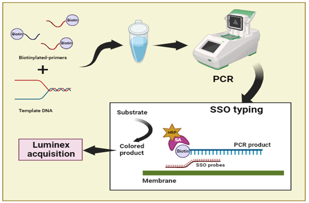
HLA SSO typing was carried out on the basis of hybridization of SSO probe with
single stranded PCR product. In this technique, the HLA-A and HLA-DR locus specific biotinylated-primers were used to amplify the HLA loci and then the obtained product was placed on the microsphere containing SSO probes. Each probe was designed to hybridize with its complementary region which may be present in amplified DNA. After washing, only strands complementary to probes were attached on membrane (Figure 1). Then streptavidin was added to the membrane which reacted with biotin and Luminex instrument recorded the fluorescent which detect the different gene of HLA. For performing Luminex assay, instrument was switched on 30 minutes before conducting and sample was protected from light. After completion of batch, data was exported as Comma Separated Values (CSV) file. This method was performed by using the protocol given by [27]. Results generated by SSO typing can be used to analyze the possibility of presence of all alleles present in sample and also the sequence of interest in amplified DNA.
Figure 1: Sequence-Specific Oligonucleotide (SSO) typing, Amplification of template DNA through biotinylated-primers followed hybridization using SSO probes followed by Luminex acquisition
Match it DNA Software
The typing of samples were performed by using different spread sheet programs including Microsoft Excel, Graphpad prism 6 etc. according to available data, the minimum number for every SSO sample was verified and values of consensus probes were determined which should be above than minimum median fluorescent intensity.
RESULTS
The collected data were composed of 96 % individuals were Punjabi, 2 % Saraiki and 2 % Pukhtun. In case of blood groups, individuals with blood groups 30 % A+, 1 % A-, 81 % B+, 2 % B- and 12 % AB+ were analyzed and there were 128 % males with ratio of 73 % females. The age groups 30-36 and 37-43 were selected for locus analysis.
Frequency of HLA-A Locus in Different Individuals
Different Blood Groups
The individuals having with blood group A, B, AB, O was compared on the basis of HLA-A loci. The individuals with different blood group had different allele for HLA. Figure 2 (a) represents the frequency of allele 1 of HLA-A locus which were high for blood group B+ while least for blood group O-, B- and A-, (b) the allele 02 was compared in different blood groups, O+ blood group showed maximum frequency as compared to A+ and AB+, (c) represents more frequent number of allele 3 for blood groups O+ and B+. Similarly, blood group O+ was more frequent in locus 11, 23, allele 29, 33 and in locus 24 (Figure 2 d, e, f). In individuals having HLA allele 26, blood groups A+, B+ and AB+ were more frequent while allele 68 was frequent in blood Group O+ and A-. Maximum results were obtained in case of allele 23, in which number of individuals exceeds up to 20-25%.
Donor and Recipient
The HLA typing was performed on the basis of donor and recipient allele frequency. The allelic frequency of donor showed that the 11 were more frequent in all donor as compared to other alleles while allele 1 and allele 2 were more prevalent in all recipients while comparing to rest of allele 3, 11, 24,26,29,23 and 68 (Figure 3).
Different Ethnic Groups
The individuals’ belonged to different ethnicity had variety of allelic frequency as Pukhtun with locus 3 showed frequent individuals and there were no participants with allele 1 and 2 present in Pukhtun group. Among different ethnic groups, similar frequency was observed in the cases of Punjabi and Saraiki with allele 68, in which individuals exceeded up to 60-80%. The individuals’ belonged to Punjabi had less frequency for allele 1, 2, and 3 while Pukhtun group was less frequent to 3-4 % (Figure 4).
Different Gender
The frequency of allele 68 showed maximum results in both male and female population while male and female allele 01 showed least frequencies limited to 0.5%. Maximum result was obtained in between 60 to 80% in case of allele 68 (Figure 5).
Different Cities
Figure 6 (a) represent the high allelic frequency of allele 1 among Gujranwala population, the individuals from Lahore showed high allelic frequency of allele 2, 3 and 33 (Figure 6 b, c, i) while in Faisalabad, the individuals had high frequency number of allele 26, 29, and 33 (Figure 6 g, h, i). The other cities were also showed frequent number of alleles among their population i.e. allele 3 in Parachnar, allele 11 and 29 in Kasur, allele 23 in Sahiwal, allele 24 in Sialkot, allele 29 in Narowal, allele 68 in Bhakkar and Okara. The maximum frequency was observed in Sahiwal population in range 20-25 %.
Different Age Groups
The individuals were distributed among different groups on the basis of different age number. Figure 7 showed variety of results of HLA-A loci according to different age groups. The alleles 1, 3, 11, and 26 were frequent in the individuals of age group 23-29 while alleles 24, 26, 29, and 33 were more prevalent among age group 30-36. The allele 2 in 44-50, allele 24 in 16-22, and allele 23 were observed among individuals of less than 50 years. Some age groups showed less frequent results of allelic frequency i.e. 37-50 etc (Figure 7).
Frequency of HLA-DR Locus in Different Individuals
Different Blood Groups
The allele frequency of different alleles (allele 1, 3, 4, 7, 8, 9, 10, 11, 12, 13, 14, 15) of HLA-DR was determined among different blood groups. The high frequency number of allele 1, 3, 7, 8, 9, 10, 12, 13, and 14 was observed among individuals of blood group O+ while allele 4 was frequent among individuals of blood group A+. The blood group B+ individuals showed maximum allelic frequency of allele 11 and 15 (Figure 8).
Donor and Recipient
The highest allelic frequency of allele 3 was observed in donor group while allele 2 and 3 was highest among recipients. Peak frequencies were obtained in the case of donor in whom number of individuals exceeds up to 20 to 30% (Figure 9).
Different Ethnic Groups
Among different ethnic groups, the allele 3 was frequent in Punjabi and Saraiki while
allele 7 and allele 9 were more recurrent among Pukhtun population. Punjabi showed peak frequencies with 30-40% individuals as compared to Pukhtun in which percentage was in between 1 to 1.5% (Figure 10).
Different Gende
Figure 11 showed the maximum frequency of different alleles of HLA-DR between male and female. The allele 3 showed more prominent results in both male and female groups while the rest of alleles were least frequent between male and female distribution (Figure 11).
Different Age Groups
The HLA-DR alleles were determined among different individuals also on the basis of different age groups. The allele 1, 3, 8, and 10 was recurrent among the age groups 37-43 while allele 3, 4, 7, 9, 10, 11, 12, and 14 were frequent among individuals of age 23-43. The allele 3, 8, and 14 was also observed in age group 30-36. The individuals of age group 16-22 showed maximum frequency for allele 8, 10, 14, and 15 while allele 13 was observed in approximately all age groups (Figure 12).
Different Cities
Figure 13 showed the allelic frequency of HLA-DR among individuals of different cities of Pakistan. The allele 1 and 10 was frequent in Kuchai while allele 3 and 11 was observed among the individuals of Kasur. Similarly other alleles i.e. allele 4 in Sialkot, allele 7 in Lahore and Sheikhupura, allele 8, 10 and 13 in Faisalabad, allele 9 in Parachnar, allele 10 also observed in Pakpatan and Vehari, allele 11 and 12 in Gujranwala, allele 13 in Gujrat, Lahore and Vehari, allele 14 in Rajanpur and allele 15 was observed in Lahore (Figure 13).
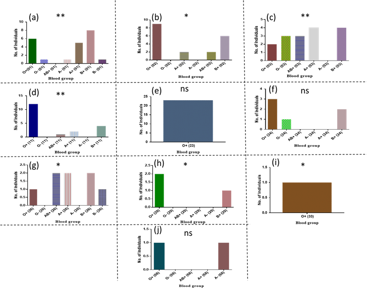
Figure 2: Frequency of different allele of HLA-A with respect to different Blood groups, (a) Locus 01, (b) locus 02, (c) locus 03, (d) locus 11, (e) locus 23, (f) locus 24, (g) locus 26, (h) locus 29, (i) locus 33, (j) locus 68
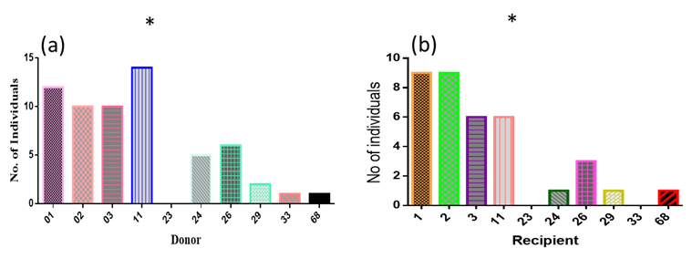
Figure 3: Frequency of different allele of HLA-A with respect to Donor and recipient (a) Donor (b) Recipient
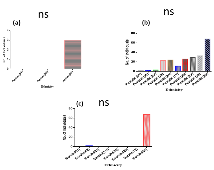
Figure 4: Frequency of different allele of HLA-A with respect to ethnic groups. (a) Pukhtun (b) Punjabi (c) Saraiki
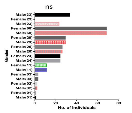
Figure 5: Frequency of different alleles of HLA-A with respect to Gender
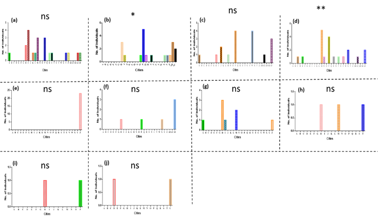
Figure 6: Frequency of different alleles of HLA-A with respect to Cities, (A)AliPurChatha (B)Bhawalnagar (C)Bhawalpur (D)Bhakkar (E)Shahdra (F)DeraGaziKhan (G)Gojra (H)Faisalabad (I)Gujranwala (J)Gujrat (K)Jhang (L)Jehlum (M)Kasur (N)Khanewal (O)Kuchai (P)Lahore (Q)Laiyah (R)MuzafarGarh (S)NankanaSahib (T)Narowal (U)Okara (V)Pakpatan (W)Parachnar (X)Rajanpur (Y)Riwind (Z)Sahiwal (AB)Sargodha (AC)Sheikhupura (AD)Sialkot (AE)Vehali (AF)Vehari
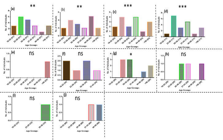
Figure 7: Frequency of different alleles of HLA-A based on Age Groups. (a) locus 01 (b) locus 02 (c) locus 03 (d) locus 11 (e) locus 23 (f) locus 24 (g) locus 26 (h) locus 29 (i) locus 33 (j) locus 68
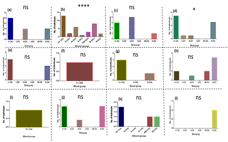
Figure 8: Frequency of different alleles of HLA-DR with respect to blood groups (a) locus 01 (b) locus 03 (c) locus 04 (d) locus 07 (e)locus 08 (f) locus 09 (g) locus 10 (h) locus 11 (i) locus (12) (j) locus 13 (k) locus 14 (l) locus 15
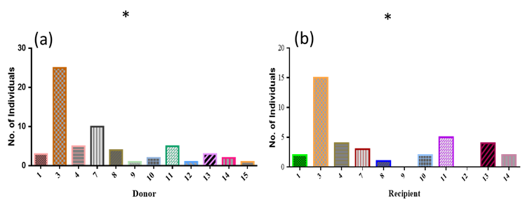
Figure 9: Frequency of different alleles of HLA-DR with respect to donor and recipient (a) Donor (b) Recipient
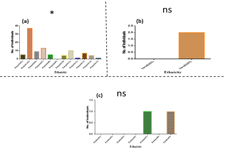
Figure 10: Frequency of different alleles of HLA-DR with respect to Ethnic Groups (a) Punjabi (b) Saraiki (c) Pukhtun
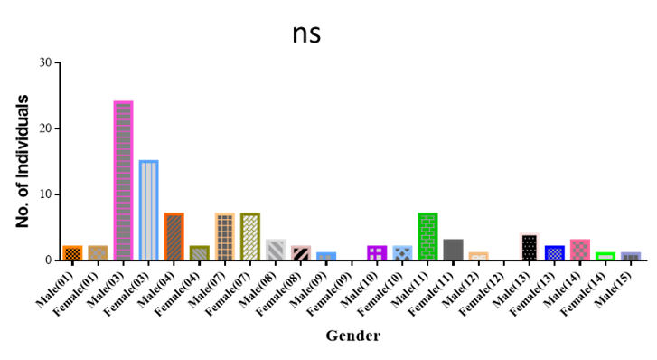
Figure 11: Frequency of different allele of HLA-DR with respect to Gender
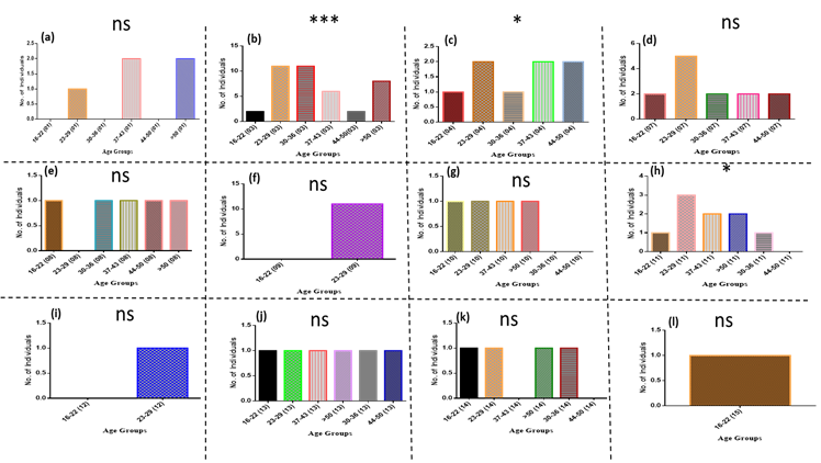
Figure 12: Frequency of different alleles of HLA-DR with respect to Age Groups, (a) locus 01 (b) locus 03 (c) locus 04 (d) locus 07 (e) locus 08 (f) locus 09 (g) locus 10 (h) locus 11 (i) locus (12) (j) locus 13 (k) locus 14 (l) locus 15
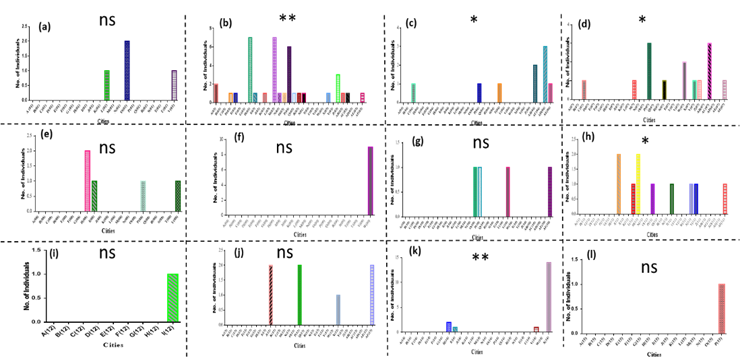
Figure 13: Frequency of different alleles of HLA-DR with respect to Cities, (A)AliPurChatha (B)Bhawalnagar (C)Bhawalpur (D)Bhakkar (E)Shahdra (F)DeraGaziKhan (G)Gojra (H)Faisalabad (I)Gujranwala (J)Gujrat (K)Jhang (L)Jehlum (M)Kasur (N)Khanewal (O)Kuchai (P)Lahore (Q)Laiyah (R)MuzafarGarh (S)NankanaSahib (T)Narowal (U)Okara (V)Pakpatan (W)Parachnar (X)Rajanpur (Y)Riwind (Z)Sahiwal (AB)Sargodha (AC)Sheikhupura (AD)Sialkot (AE)Vehali (AF)Vehari
DISCUSSION
The human leukocyte antigen (HLA) is a major class of major histocompatibility complex (MHC) and their genes are commonly present on chromosome 6p21 [28]. These are the most diverse group of antigens and contain polymorphism even in a single population. These were discovered by observation of antigenic difference in white blood cells taken from different individuals. There are two major classes of MHC i.e. class I and class II, there is another class III but with less importance as class III has no well defined role as compared to class I and class II. Class-I has three loci including HLA-A, HLA-B and HLA-C while class-II has five loci including HLA-DR, HLA-DQ, HLA-DP, HLA-DO and HLA-DM. The HLA genes are considered more polymorphic and they show co-dominance in their expression [29]. The HLA typing is very important step in forensic sciences on the basis of detection of sequence polymorphism. It can be done through Sequence-Specific Oligonucleotide (SSO) and Allele-Specific Oligonucleotide (ASO) typing. Genetic identity testing is used to determine the unique characteristics of individual’s genome. It is being recorded that almost all the human genome is identical but there is relatively less number of different genes are present. The most common example of this uniqueness is HLA genes as they carry polymorphism to great extent. Some studies have reported that there are approximately 1250 different alleles of HLA (human lymphocyte antigen). The major function of HLA protein is to present pathogenic peptides on the surface of T cells which further treated by adaptive response of T cells. The HLA class usually belongs to the groups of molecules called as Immunoglobulin superfamily. The HLA molecule with foreign peptide presenting on it first recognize by T cell receptors and then T cells differentiate between self and non-self-peptides.
This study was based on HLA-A and HLA-DR typing by using SSO typing method. HLA-A express on nucleated cells and involved in identification of infected cell and destroy it with cytotoxic T cell. While class-II HLA-DR express on Antigen presenting cell (APC), B lymphocytes and active T lymphocytes and identify foreign antigen with the help of helper T cell. Leen and co-workers performed an experiment through Antony Nolan register based in United Kingdom. They collected the HLA frequency data from different asian countries including Bangladesh, India, Pakistan, and East Asian countries and made a comparison between the frequencies of different alleles of HLA genes. They have reported that the most frequently occurring haplotype of HLA-A was allele 1 in any population and second most common allele was allele 33 among different populations [28].
The current study has shown that HLA-A 68 was the most common haplotype observed in Pakistani population from different cities. It shows the diverse nature of HLA-A haplotype. It is continuously changing according to their needs. Similarly another experiment was performed by Esmaeili and his team in which they determined the frequencies of HLA-A haplotype among the population living in a city of Mashhad, Northeastern Iran. They have reported that the HLA-A 2 allele was the most frequent haplotype among this population while in comparison to Pakistan (as in current study) allele 68 was the most frequent [29]. It showed that HLA-A haplotypes are also different for different parts of the world. Dere et al., 2020 conducted an experiment in eastern of Turkey in which they determined the frequencies of HLA-DR polymorphism and they reported that there are different sub groups of HLA-DR and the most common haplotype was HLA-DRB1*4 and HLA-DRB1*14 among the patients of Pemphigus vulgaris virus [30] while in comparison to current study, HLA-DR had observed with different perspectives in population including blood groups, age, gender, ethnicity and cities.
CONCLUSION
By interpreting analysis of blood groups, cities, gender, age and ethnicity of Pakistan, it has been observed different alleles of human leukocyte antigen have different proportion even in the same local population of Pakistan. In this study, different locus responsible for polymorphism and hence kidney failure is highlighted. It is crucial to study HLA polymorphism for complete eradication of transplant rejection and better therapy response in local population. In Pakistan, the study of HLA polymorphism is of great significance as alleles encoded by HLA is highly polymorphic so differently tuned adaptive immunity. Among all HLA-A haplotype, locus 68 showed maximum frequency with respect to different perspectives while HLA-DR showed diverse nature of results with different proportion. To avoid rejection by polymorphism, HLA matching is important to engraft donor cells efficiently and to avoid any complication of graft versus host disease (GVHD).
REFERENCES
*Correspondence: rabbiajawad000@gmail.com
[1]Department of Life Sciences, University of Management and Technology, Lahore, Pakistan
[2]School of Biological Sciences, University of the Punjab, Lahore, Pakistan
[3]Undiagnosed Rare Disease Clinic, Department of Medical and Molecular Genetics, Indiana University School of Medicine, Indiana, USA