Detection and Characterization of Antibiotic-Resistant Microorganisms in Cosmetic Products
Aneeba Fatima1 | Rida Batool1 | Nazia Jamil1*
1Institute of Microbiology and Molecular Genetics, University of the Punjab, Lahore. Pakistan
*Correspondence
Nazia Jamil
Email: nazia.mmg@pu.edu.pk
ABSTRACTThis study aimed to determine the prevalence and antibiotic resistance among the bacteria isolated from the makeup products of two salons. This study has significant importance as it raises the overlooked issue of antibiotic resistance in cosmetic products and the mode of transmission of antibiotic resistance from one person to another by using shared make-up products in salons. 20 samples of all the make-up items (face powder, primers, contour, blush-on, lipsticks, lip-gloss, eye-liner, eye-shadow, and mascara) were collected from salons in Lahore and Shakargarh, Pakistan. The microbial load reported in samples exceeded the threshold limit set by the Scientific Committee on Consumer Safety, and Food and Drug Administration Authority. Then, the antibiotic resistance of all strains was assessed using the different stock concentrations and Muller-Hinton Agar. The highest resistance of 14.29% was reported against fluoroquinolone,11.90% for Tetracycline,9.52% for chloramphenicol, 2.38% to Gentamycin, and no resistance was observed against neomycin and streptomycin. From all biochemical tests, the tentatively identified strains include Staphylococcus aureus, Shigella sp., Enterococcus sp., Neisseria sp., Pseudomonas aeruginosa, Bacillus sp., and Citrobacter sp. Among the antibiotic-resistant strains, the IntI 1 gene was detected in two strains, then these strains were identified by the Sanger sequencing method and these strains included Acinetobacter shindleri with accession number PP978765 and Enterobacter cloacae with accession number PP978766. From the results, it can also be concluded that these strains exhibit IntI 1 gene-mediated resistance. There is an urgent need to tackle the growing issue of resistance to cosmetic products. This study highlights the importance of raising awareness among people and calls for stricter hygiene regulations in salons and the cosmetic industry to ensure safety and quality. Keywords: Antibiotic Resistance, Cosmetic Products, Microbial Contamination, Integron Genes, Heavy Metals |
The term cosmetic encompasses a lot of articles with a variety of purposes. Cosmetic products modulate physical aesthetics and augment the human body attractiveness [1]. Makeup is one of the the main component included in the category of cosmetics. Makeup products have played a significant role in reducing stress among women by helping them conceal imperfections and enhance their appearance [2]. Thus, the beauty salon industry has been progressing rapidly in recent years [3]. In beauty salons, the microbiological risks associated with infections vary depending on the different levels of hygiene procedures implemented by the salons [4]. In many local salons across Pakistan, the same makeup products are used on multiple clients, which exposes the products to different people’s skin and can lead to contamination with skin bacteria. This practice increases the risk of spreading infections through shared makeup products, brushes, and applicators [4].
Makeup products can provide an ideal environment for microorganisms to grow due to their composition. These products often contain ingredients like minerals, sugars, salts, moisturizers, colors, preservatives, and other organic or inorganic compounds. Combined with their moisture content, they create favorable conditions for microbial contamination [5]. Contamination typically occurs in two main ways: first, during production or packaging if proper hygiene isn’t maintained, and second, during use—particularly in salons where the same products are shared among multiple clients. Additionally, tools like brushes and applicators used repeatedly in salons further increase the risk of spreading contamination [6].
Different studies have shown that the high prevalence of microbes in make-up products causes several severe health problems, especially in immunocompromised patients. These micro-organisms frequently report contamination in makeup products and cause a variety of health-related problems: Staphylococcus epidermidis, Staphylococcus aureus, Escherichia coli, Klebsiella pneumonia, Enterobacter sp., pseudomonas aeruginosa, Aspergillus niger, Micrococcus sp., Bacillus sp., Acinetobacter sp., Citrobacter sp., Candida albicans, and various other Aspergillus species [7]. The major commonly found microbes that pose a major threat to consumers' health include Pseudomonas aeruginosa, Staphylococcus aureus, Candida albicans [7], and Aspergillus brasiliensis. These are also restricted by EU pharmacopeia for their small contamination, even 1g/ml, in cosmetics and constituents of make-up items [8].
The health risks linked to using contaminated cosmetics can vary depending on the product, the type of service, and the tools used. Skin infections are the most common, often caused by contaminated products like face powders, foundations, mascaras, and eyeliners. These infections, which can include conditions like acne, boils, bullous impetigo, and scalded skin syndrome, are most commonly caused by Staphylococcus aureus. [7,8] and S. epidermidis [7]. A wide range of cases of allergic contact dermatitis was reported in some regions of the Middle East. Herpes causes blisters on the lips and around the mouth through the use of shared lipsticks. The shared lipsticks and make-up brushes also serve as a source of spreading allergic contact dermatitis to other people [9]. Another study concluded that Pseudomonas aeruginosa and Serratia liquifaciens are the potential sources of eye infections, itching, pink eye, corneal ulcer, and conjunctivitis and if conditions become severe can turn into blindness [8]. Metabolites and endotoxins produced by the micro-organisms can cause irritation, itching, acne, eye damage, cancer, and other health conditions. It has also been reported that S. sciuri caused wound infection, peritonitis, septic shock, and endocarditis [7].
Microorganisms isolated from cosmetic products have shown resistance to various antibiotics, which may develop through mechanisms such as integron-mediated resistance, efflux pumps, and biofilm formation. As antibiotic resistance continues to rise at an alarming rate, integrons play a significant role in its rapid spread. Studies have shown that integrons are genetic elements responsible for conferring multi-drug resistance, particularly in Gram-negative pathogens [10]. These integrons can acquire antibiotic resistance genes from the environment and transfer them to other bacterial hosts, thus contributing to the spread of antimicrobial resistance [11]. Class I integrons, in particular, contain a gene called IntI1, which indicates the transfer of antibiotic-resistance genes among bacteria. [12]. In addition to microbial contamination, beauty products can also contain harmful substances like aromatic compounds, heavy metals (such as mercury and chromium), volatile organic compounds, and particulate matter, all of which can be extremely harmful to consumers' health [13].
This study is focused on the isolation and identification of antibiotic-resistant bacteria from make-up products of salons, evaluating their resistance to heavy metals, and the 16S rRNA sequencing of highly resistant strains.
Samples of all make-up products (foundation, lipstick, face powder, Blush-on, contour, lip gloss, mascara, eye-liner, and eye-shadow) were collected from 2 salons, Salon A of Johar town, Lahore, and Salon B of Shakargarh in November 2023. Both urban and rural settings were considered when selecting the samples to be collected from salons in Lahore and Shakargarh in order to have a more general idea regarding the use of makeup products and the presence of microbes on those products in different areas. Sample transport was done carefully in transport media by following the aseptic conditions and SOPs. The expiry date of the samples was considered during collection and none of the samples was expired [14].
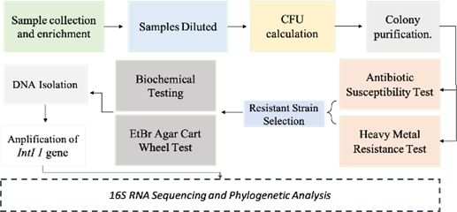
2.2 Study Design
Figure 1. Flowchart of the methodology of study. The figure explaining all the procedures performed in the study step-wise
2.3 Sample Processing and Isolation of Microbes
Sample collection containers were observed for any kind of leakage or irregularities. Sample content was removed from containers by disinfecting the surface with 70% ethanol. For sample enrichment, Media L-broth was prepared. After media autoclaving, all the samples according to their texture, were processed differently and were aseptically added to L-broth test tubes and then incubated on a shaking incubator at 37°C for 18-24 hours. After sample enrichment, the dilution method was performed and dilutions of samples were prepared from 10₋1 to 10₋8and L-Agar and MacConkey media were prepared to cultivate bacteria of sample make-up products.
50µl of 10₋1 and 10₋3 dilutions of all samples were spread properly with the help of a sterile spreader on autoclaved overnight L-agar and MacConkey plates. Plates were incubated at 37°C for 18-24 hours. After 18-24 hours, results were visualized and a variety of flora was observed on plates, then their CFU was calculated by counting the no. of colonies [14]. This formula was used for calculating the CFU/ml of all the samples:
2.4 Antibiotic Susceptibility Test
This test was performed to check the sensitivity or resistance of microbes against the antibiotics. A total of 42 strains were isolated from different samples and were tested for antibiotic sensitivity. This was done using two methods: Stock solution of antibiotics and Kirby Bauer disk diffusion method.
Tetracycline and Chloramphenicol were selected for the first process at 2 different concentrations. The stock solution of the first antibiotic Tetracycline was made in ethanol at a concentration of 10mg/ml and then their working solutions were prepared at 2 concentrations of 50µg/ml & 100µg/ml and the stock solution of 2nd antibiotic chloramphenicol was made in autoclaved distilled water at a concentration of 10mg/ml. Then, their working solutions were prepared at 2 concentrations of 100µg/ml &500µg/ml. Their working solution was mixed in slightly warm autoclaved agar and poured into the plates. All the strains were streaked over the poured plates and incubated for 24 hours at 37°C to check the antibiotic sensitivity or resistance. After 24 hours, results of strain growth over antibiotic-containing media were observed and noted.
For the second method, the Kirby-Bauer Disk Diffusion protocol was followed [15]. For this, the medium of choice was Muller-Hinton agar. So, after pouring and solidification of media, a sterile cotton swab was used to make a lawn of inoculum to ensure confluent growth. Then, filter paper discs containing the specified concentration of 6 different antibiotics are placed over the surface of Muller-Hinton agar which is seeded with the lawn of bacteria for whom resistance or sensitivity needs to be checked. Then the plates are incubated at 37°C for 24 hours. The result was observed by checking the presence or absence of zones of inhibition and by measuring their diameter. Thus, by measuring the size of the zone, we can assess the susceptibility of the microbe to the specific drug [15].
The resistance pattern for these six antibiotics is then analyzed by using statistical analysis. Chi-square was applied overall data of the antibiotic resistance profile to check whether the distribution of resistance of these six antibiotics vary from each other or not.
2.5 Heavy Metal Resistance Test Among Isolated Strains
Heavy metals may be present in the make-up products and can be hazardous for human health so, this test was performed to check whether the microbes isolated from make-up products can grow in the presence of heavy metals or not. For this purpose, 2 heavy metals Dichromate and Mercury sulfate are used at 3 different concentrations of stock i.e at 50mM, 25mM, and 5mM to check at which concentration bacteria will be able to grow over the heavy metal, whether they are resistant to all concentration or sensitive. For performing this practical, autoclaved N-agar was mixed with 1mM working solutions of 3 different concentrations of both heavy metals. Heavy metal solution was properly mixed in the warm agar by swirling. Then, it was streaked with all 42 strains and incubated for 24 hours at 37°C. The next day's result was observed by checking whether growth had occurred or not [14].
2.6 Biochemical Tests
Biochemical tests of selected strains were performed for the further identification of genus and species of bacteria. These tests were performed by following the standard protocol given in the laboratory manual. These tests include Gram-Staining, Catalase, Oxidase, Spore staining, Starch hydrolysis, Citrate test, Methyl Red test, VP test, and Triple Sugar Iron test. The results of these tests helped in the tentative determination of highly resistant strains [14].
2.7 EtBr Agar Cartwheel Method
EtBr Agar Cart Wheel Method was performed to check the efflux-mediated multidrug resistance in the bacteria. To perform this test, the selected strains were streaked in the cartwheel pattern over the solid media that was seeded with different concentrations of EtBr to detect the efflux activity of different and whether they were capable of extruding the EtBr from the cell or not. The fluorescence emitted will be observed under UV and this fluorescence has an inverse relationship with the capacity to extrude the EtBr [16].
2.8 Isolation and Analysis of Genomic DNA
Nucleic acid extraction in highly resistant strains was performed with the help of the mini-DNA Extraction kit using a Thermo Scientific GeneJET Genomic DNA purification kit. The isolated genomic DNA was then analyzed by gel electrophoresis using 1X TAE Buffer and 1% agarose gel [17].
2.9 PCR Amplification of IntI1Gene
PCR amplification of the IntI1 gene was done and sterilized conditions were maintained for the preparation of the PCR reaction mixture. The program was set at different temperatures for different steps and different times for different steps i.e initial denaturation at 95°C (5 min), final denaturation at 95°C (45 sec), annealing at 60°C (35 sec), extension at 72°C (1 min), and final extension at 72°C (10 min). Thirty cycles were set for this program [18].
The sequence of the forward primer of the IntI 1 gene used was 5’CCTCCCGCACGATGATC3’ and the sequence of the reverse primer was 3’TCCACGCATCGTCAGGC5’.
2.10 16S rRNA Gene Sequencing and Phylogenetic Analysis
Two highly resistant strains PD2 and PD4 in which the IntI 1 gene was present were selected and cultivated on a nutrient agar plate and 16S rRNA sequencing was done by Sanger dideoxy sequencing. Sequences were checked for their maximum homology against GenBank using BlastN. After the sequencing, phylogenetic trees of strains were constructed using MEGA 4.
Enumeration of Sample Microbial Flora on Luria Bertani Agar Plates:
50ul of each dilution was spread on LB and Mcckonkey agar plates by following the spread plate method. The results obtained from the spreading of plates are given in supplementary data Table 1.
Antibiotic resistance of all strains was firstly checked by the stock concentration of two antibiotics Tetracycline and Chloramphenicol and the results are given below in table 2.
Antibiotic resistance was then determined over the Muller-Hinton Agar and it was found that 90.48% strains showed sensitivity to Chloramphenicol while only 9.50% of strains were resistant to Chloramphenicol. Similarly, among all strains only 2.38% strains showed resistance to gentamycin and the remaining were sensitive to it. Among the strains, isolated from all the samples of makeup products,85.71% were found sensitive to fluoroquinolone while 14.29% were found resistant to fluoroquinolone. For the Tetracycline,11.90% of strains showed resistance while all other was found sensitive to it and for streptomycin, no strain were found resistant.
Table 1. Total CFU/ml found in samples and Product contamination range
|
Product Type |
No. of samples |
No. of contaminated samples |
Threshold of contamination allowed: |
||
|
e CFU/g |
|||||
|
< 103 |
103–105 |
> 105 |
|||
|
Foundation |
2 |
0 |
0 |
2 |
<103 CFU/g |
|
Primer |
2 |
0 |
0 |
2 |
|
|
Powder |
2 |
0 |
0 |
2 |
|
|
Contour |
2 |
2 |
0 |
0 |
<103 CFU/g |
|
Lipstick |
2 |
0 |
1 |
1 |
<103 CFU/g |
|
Lip gloss |
2 |
0 |
1 |
1 |
<103 CFU/g |
|
Blush-on |
2 |
0 |
0 |
2 |
<103 CFU/g |
|
Mascara |
2 |
0 |
1 |
1 |
<102 CFU/g |
|
Eye-Liner |
2 |
0 |
0 |
2 |
<102 CFU/g |
|
Eye-Shadow |
2 |
0 |
0 |
2 |
<102 CFU/g |
Table 2. Antibiotic Resistance profile over stock concentration of antibiotics
|
Resistant Strains |
|||
|
Tetracycline 50µg/ml |
Tetracycline 100µg/ml |
Chloramphenicol 100µg/ml |
Chloramphenicol 500µg/ml |
|
LG2 |
LG2 |
LS4 |
F2 |
|
B2 |
M1 |
M1 |
LS4 |
|
ES1 |
ES1 |
PD2 |
LG2 |
|
ES2 |
ES2 |
PD4 |
LG4 |
|
PR3 |
|
PR3, PR4 |
PR3 |
|
LS1 |
|
F2 |
PD4 |
|
M1 |
|
ES4,CN4 |
|
|
PD5 |
|
PD1 |
|
|
PR3 |
|
LG2 |
|
|
PR4 |
|
LG4,LG5 |
|
|
|
|
M1,M2,M3 |
|
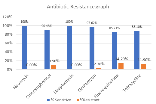
Figure 2. Chart representing the Antibiotic Resistance and Sensitivity Pattern among Isolated Strains. This bar graph illustrates the percentage of antibiotic-sensitive and resistant bacterial strains for six antibiotics. The blue bars represent sensitive strains and the orange bars represent the resistant strains. Most importantly, there was no response to Neomycin and Streptomycin, and maximum resistance was recorded to Fluoroquinolone (14.29% and Tetracycline (11.90%).
The resistance pattern of six antibiotics was tested by applying the chi-square test in SPSS version 25 to test the significance of the differences in the resistance distribution of the antibiotics. The chi-square test was performed by comparing the observed frequencies of resistance against six antibiotics to the expected frequency which was derived from the hypothesis of no difference in resistance frequency.
3.3 Hypothesis Tested
Table 3. Chi-Square Analysis Results
|
Table value of chi-square |
Critical value(α=0.05) |
Degree of freedom |
Calculated value of chi-square |
p-Value: |
|
11.07 |
0.05 |
5 |
11.71 |
0.039 |
As the p-value found is 0.039 and is less than the critical value of 0.05, Thus, we reject the null hypothesis. This demonstrates a significant difference in the dispersion of the resistance between the six antibiotics, which means that the study found out that some of the antibiotics have higher levels of resistance compared to others. Further, it was seen that antibiotics such as Fluoroquinolone and Tetracycline showed prominent resistance in comparison with Neomycin and Streptomycin resistance.
It is established from these observations that bacteria may or may not be sensitive to antibiotics and there is a strong need to consider rational antibiotic usage. Besides, they emphasize the need to have effective measures to check microbial infiltration in cosmetics for safety and to avoid the effects of antibiotic resistance among consumers.
Heavy metal resistance of all strains was checked and no strain was observed in the presence of heavy metal in the agar plates. Thus, almost all strains were sensitive to Heavy metals.
Out of 42 strains,10 strains were selected that were resistant in the presence of both stock concentration and antibiotic disk or at least they were showing resistance to 2 antibiotics in the Antibiotic-disk test. These are mentioned below in table 4.
The biochemical characterization of the selected resistant strains was done and their tentative classification was performed.
Table 4. Selected Antibiotic-Resistant Strains and their tentative classification
|
Strain Names |
No. of antibiotics resistance to (Out of total 6) |
Biochemically Identified strain name |
|
F2 |
3 |
Staphylococcus aureus |
|
PD2 |
3 |
Acinetobacter shindleri |
|
PD4 |
3 |
Enterobacter cloacae |
|
PR3 |
2 |
Pseudomonas aeruginosa |
|
PR4 |
2 |
Neisseria sp. |
|
LS4 |
2 |
Citrobacter sp. |
|
LG2 |
3 |
Enterococcus sp. |
|
LG4 |
2 |
Shigella sp. |
|
M1 |
3 |
Bacillus sp. |
|
CN4 |
2 |
Enterobacter sp. |
3.4.2 Screening of IntI1 Gene by PCR
PCR amplification of the IntI1 gene of two resistant strains was done to check the presence of the IntI1 gene in the resistant strains and it was found that band formation of the PD2 strain (1200 bp) and PD4 strain (1000bp) appeared over the PCR Gel. So, the strains PD2 and PD4 were found positive for IntI1 gene detection.
The accession number of sequences submitted to GenBank is recorded below in Table 5.
The evolutionary Relationship of Strain PD2 with related taxa is given below in Figure 3.
The evolutionary Relationship of Strain PD4 with related taxa is given below in Figure 4.
So, the identified antibiotic-resistant strains of our study were two strains. PD2 is identified as Acinetobacter shindleri and the accession number is PP978765. Second is strain PD4 named Enterobacter cloacae and the accession number is PP978766.
Both these strains were detected positive for the IntI 1 gene, thus the antibiotic resistance in these strains may be IntI 1 gene-mediated resistance.
Table 5. Accession numbers of strains identified by 16S rRNA gene sequences
|
Accession numbers of 2 Highly Resistant Strains. |
|||
|
Strain name |
Organism name |
Sequence Length(bp) |
Accession Number |
|
PD2 |
Acinetobacter shindleri |
1160 |
PP978765 |
|
PD4 |
Enterobacter cloacae |
327 |
PP978766 |
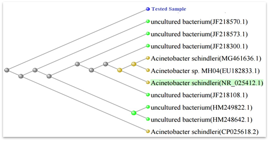
Figure 3. Phylogenetic Tree of Acinetobacter schindleri.it shows the position of the tested sample of NMA in the genus, family, and order. The phylogenetic tree compares the tested sample with other bacterial species: there is a closer match with the *Acinetobacter schindleri* (NR_025412.1) and some other unidentified bacterial strains.
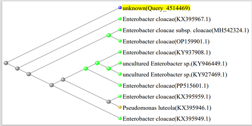
Figure 4. Phylogenetic tree of Enterobacter cloacae. Nucleotide sequence neighbor-joining tree based on 16S rRNA gene sequence comparing the unknown query sequence (Query_4514469) with *Enterobacter cloacae* and related species. The tree depicts the indication of close clustering with *Enterobacter cloacae* (for example KX395967.1; OP159901.1; KY937908.1) as well as with subspecies *Enterobacter cloacae subsp. cloacae* (MH542324.1) and the uncultured *Enterobacter sp.* (KY946449.1; KY927469.1).
Makeup products are a part of many people’s daily routines, helping them feel confident and enhance their appearance. However, sharing these products and tools can lead to the spread of antibiotic-resistant bacteria, which is a growing concern. [9]. The European Scientific Committee on Consumer Safety (SCCS) recommends that makeup products for sensitive areas, like the eyes or mucous membranes, should have a microbial load below 100 CFU/ml. For other types of makeup, the limit is 1000 CFU/ml. Unfortunately, our study found that many eye and general makeup products exceeded these safety limits, raising concerns about their safety [19]. In the eye-shadow sample, the microbial count found during our study by spreading method was 693000 CFU/ml in one sample and it varies in other samples but it also crossed the limit set by SCCS. In mascara, the microbial count reported superseded the limit [19]. In a study conducted [7], the microbial load of aerobic mesophilic bacteria in mascara was 3.6×10³cfu/ml while in our study the load reported was higher [7]. The reason was that we used the highly used sample of the salon.
Moreover, the US FDA has also set some standards and microbiological limits over the make-up products and these vary from the SCCS. According to the US FDA, the set limits for cosmetics are 500 CFU/ml or per gram while the threshold limit for all other products is ≤ 1000cfu/g or ml. However, in our study contamination in all the products exceeded the mentioned limits of both SCCS and US FDA [19].
For the other product, e.g. face powder the microbial load reported in our study was 16×106 CFU/ml in one sample and 4.86×10⁶ CFU/ml in another sample and it was far beyond the limit set by SCCS and US FDA. The microbial load reported in our samples collected from two different salons varies but the limit remains superseded from the standard limit [19] According to a study conducted [7], the total number of bacterial counts found in lipstick samples corresponds with the load isolated from our samples with little variations. Our results correspond similarly to the findings of another study conducted by Noor et.al that reported that beauty shops and salons act as vehicles of infection among users [9].
Our study found that most of the tested makeup samples were contaminated with microbes, except for a few products like blush and eyeshadow. These products had lower levels of contamination because they are used less frequently and remain open for shorter periods. On the other hand, products such as face powder, lipstick, face primers, and mascara showed higher contamination levels. This was mainly due to their frequent use and longer exposure times. We identified several key factors contributing to the contamination of makeup products. These include the salon environment, which often provides ideal conditions for microbial growth, the shared use of products by multiple customers, and the prolonged usage of certain items without proper hygiene practices [2].
In our study, the bacteria isolated from make-up products of salons belong to all 4 categories according to the shape and gram reaction test. These include bacteria belonging to gram-positive rod-shaped bacteria, gram-negative rods, gram-positive cocci, and gram-negative cocci, and the strains of these categories were also found in many other studies conducted by Dimri et.al, Noor et.al, and Aslam et al. [1,2,9] In our study, the highest prevalence of gram-positive rods is 54.76%. The reason for its high prevalence is that gram-positive bacteria are mostly found in the environment so also in the salon environment. While the rate of Gram-negative bacteria found in our study was 26.19%, quite less than gram-positive bacteria. Similarly, the results obtained for gram-positive cocci and gram-negative cocci showed their very low prevalence in the make-up products as compared to the other two categories. The rate of gram-positive cocci and gram-negative cocci found is 9.52%.
The major work of our study was to find the antibiotic resistance in all the bacteria isolated from all the samples and it was seen that all strains showed sensitivity to neomycin. Enterobacter cloacae, Acinetobacter schindleri, and Enterobacteriaceae showed sensitivity to neomycin.100% strains showed sensitivity to this antibiotic as this antibiotic has a mechanism of inhibiting bacterial growth by stopping the process of translation. All the strains identified in our study were found to lack resistance mechanisms against neomycin. Similarly, all strains were sensitive to streptomycin, with no resistance genes detected. Interestingly, this aligns with findings from another study, which also reported streptomycin sensitivity in some cosmetic-related strains [20].
Among the identified strains, Acinetobacter schindleri demonstrated 100% sensitivity to neomycin, streptomycin, chloramphenicol, and gentamicin but was completely resistant to fluoroquinolones and tetracycline. Enterobacter cloacae showed resistance to tetracycline, chloramphenicol, and fluoroquinolones while remaining sensitive to other antibiotics. Similarly, the sensitivity and resistance patterns varied across other strains. For instance, Staphylococcus species exhibited 100% resistance to tetracycline and fluoroquinolones but were sensitive to gentamicin, streptomycin, chloramphenicol, and neomycin. These findings are consistent with another study, which also reported Staphylococcus species as sensitive to gentamicin [21]. Another recent work published by the University of River, Nigeria in 2023 stated the antibiotic susceptibility of microbes isolated from makeup tools i.e brushes, and it was stated that Staphylococcus aureus and Staphylococcus epidermidis were unable to grow in the presence of ciprofloxacin, levofloxacin, and gentamycin while the antibiotics whom concentration was not able to restrict the growth of bacteria was erythromycin, amoxil, norfloxacin, amplicox, streptomycin and chloramphenicol i.e showed resistance to these antibiotics [22]. Bacteria acquire this resistance by acquiring the resistant genes from resistant bacteria or by modifying the target of antibiotics. Bacillus species isolated from mascara samples showed 100% resistance to tetracycline and chloramphenicol. In a study conducted in 2022 by Kachalla et al., the antibiotic resistance reported to chloramphenicol and tetracycline was 100% which shows similarity to our research.
Statistical analysis i.e. chi-square demonstrates that the resistance distribution of all six antibiotics varies from each other. For example, strains showed high resistance to fluoroquinolones indicating the widespread transmission of fluoroquinolone resistance among microbes of salons while our data showed that less antibiotic resistance is found in the microbes isolated from make-up products of salons. Thus, resulting in varying patterns of dispersion of antibiotic resistance of all antibiotics in strains of different makeup products of the salon.
The tentative identification of our study showed the presence of Bacillus sp. in mascara samples, Staphylococcus aureus in foundation samples, Enterobacter sp. in contour samples, Shigella sp. and Enterococcus sp. in lipgloss samples, Pseudomonas aeruginosa and Neisseria sp. in the primer, and Citrobacter sp. in some lipstick samples. The strains found in our study were also reported by other studies [6]. Pseudomonas aeruginosa was also found in a study conducted by Yazici et.al in 2022 and caused a variety of diseases in the consumers. Pseudomonas aeruginosa is capable of causing eye allergy, irritation, and corneal ulcers as scratches develop on the cornea due to the use of mascara [7].
16s RNA sequencing of powder sample highly resistant strains showed the presence of Enterobacter cloacae and Acinetobacter schindleri. Enterobacter cloacae is a gram-negative bacterium and was also reported in other cosmetics products in other studies. This bacterium was also found registered in the RAPEX database in 2015-2018 as a potential contaminant of 4 different cosmetic products [6].
Acinetobacter schindleri, a gram-negative bacterium, is commonly found in the environment and acts as an opportunistic pathogen. While this bacterium has been less frequently reported in previous studies, other species of Acinetobacter have been identified in makeup products. In our study, two strains, Enterobacter cloacae and Acinetobacter schindleri, tested positive for the presence of the IntI1 gene, suggesting that their antibiotic resistance is integron-mediated.
This finding is significant, as previous data did not confirm the presence of IntI1 gene-mediated resistance in antibiotic-resistant bacteria. Our study indicates that these strains may acquire antibiotic resistance genes from their environment or skin flora through integrons such as the IntI1 gene. This mechanism facilitates the acquisition of multiple gene cassettes, including those encoding antibiotic resistance, allowing microbes to develop and spread resistance via horizontal gene transfer.
Another noteworthy observation from our study is that no microbial growth was detected under heavy metal stress. Interestingly, many studies have yet to investigate the potential for heavy metal resistance in cosmetic-associated microbial strains.
The quality and safety of makeup products used in salons are crucial factors that directly impact consumer health. Our study found that most makeup products in salons in Lahore and Shakargarh, Pakistan, showed a high prevalence of microbes. These included Enterobacter cloacae, Acinetobacter schindleri, Enterobacteriaceae, Staphylococcus aureus, Bacillus species, Enterococcus species, Pseudomonas aeruginosa, and others. The contamination levels in all samples exceeded the safety limits set by the FDA and the SCCS. To reduce the risk of infections, salons and the cosmetic industry must implement strict hygiene protocols. Regular surveillance of salons and cosmetic manufacturing facilities, along with periodic testing for microbial contamination, is essential. Additionally, staff training on contamination risks and proper hygiene practices should be prioritized to ensure consumer safety.
Statement: Authors declare no Conflict of interest
REFERENCES