Potential Enzyme Biomarkers in Clinical Diagnostics: From Theory to Practice - A Review
Atif Iqbal1 | Sikander Ali1*
1Institute of Industrial Biotechnology (IIB), GC University Lahore, Pakistan
*Correspondence
Sikander Ali
Email: dr.sikanderali@gcu.edu.pk
|
ABSTRACT Metabolism of enzymes is the basic biological process that plays an essential role in the existence of all species. As enzymes are proteins in nature, they catalyze the chemical reaction. They play a crucial role in the metabolic process of all organisms, for example: plants, animals, microorganisms, and humans, and mostly find their application in microbial technology and their diagnosis process. Any alteration or abnormality in the metabolic system of enzymes may lead to a large number of metabolic disorders. A lot of disorders associated with the components of the metabolic system of the enzyme are now used as special markers for the diagnosis of diseases in clinical studies. In this review article, we havebriefly discussed the introduction and mechanism of enzyme action with the diagnostic applications of enzymes that act as markers for disease diagnosis like it act as biosensors in disease diagnosis, used in the immune system, in the diagnosis of pathology, used in immunoassay and it may also act as a therapeutic agent in the diagnosis of disease. Keywords: Biosensors, diagnostic application, Enzyme application, therapeutic agents, immune system |
Enzymes were proteins in nature that acted as bio-catalysts and fastened the chemical reactions. Substrates were the molecules on which enzymes reacted and converted them into different molecules which were known as products. To accelerate all the metabolic events almost every cell needed enzyme catalysis that fastened the reaction necessary for maintaining life [1]. To catalyze individual steps metabolic pathways, depend upon enzymes. Enzymology is the study of enzymes and recently pseudo enzyme analysis a new field has grown up. We studied that during evolution some enzymes became disabled to function as biocatalysis, and we saw that those enzymes have unusual 'pseudo catalytic' properties and amino acids. Ribozymes were RNA molecules that showed catalytic properties. The structural specificity came in the enzyme due to its 3D structure [2].
Enzymes as catalysts can enhance the rate of reaction by decreasing the activation energy. With the use of enzymes, substrate can be converted into products more quickly than the normal reactions. Orotidine 5'-phosphate decarboxylase was an enzyme that helped the reaction to complete quickly within milliseconds and without it, this process may take millions of years to complete. Like any catalyst, enzymes were chemically the same, but they cannot alter the equilibrium of a reaction and were not consumed during the chemical reaction. However, enzymes were much more specific than most other catalysts. The activity of enzymes can be altered by invader molecules i.e.
Inhibitors were many therapeutic drugs and poisons. Outside the optimal temperature and pH, an enzyme decreased its activity markedly and when it was exposed to excessive heat, many enzymes were permanently denatured and lost their structure and catalytic properties [3]. For commercial usage enzymes were used in antibiotic synthesis. In household products, many enzymes were used to increase their chemical reactions, for example, in washing powders enzymes break down the protein, starch, or fat stained on clothes. While, in making the meat, enzymes were used to tenderize the meat so that breaks down the protein into smaller molecules [4].
Nowadays; it is quite demanding to diagnose and monitor numerous diseases for the routine assessment of clinical samples and other associated tests. To carry out clinical tests, standard analytical methods were required, which needed time collection and expert knowledge. The enzymes that are used to diagnose prognostic illness are diagnostic enzymes. Enzymes are favored for diagnosis in the presence of other proteins due to their substrate specificity and quantitative activity. It can also be employed as a tool for the detection of disease depending upon the disease severity, and it may take towards the tissue damage. Under certain circumstances, enzymes are released into the blood or plasma circulation against the diseased organ which increases the activity of enzymes [5]. Such measurement of the activity of enzymes in blood, plasma, or body fluids is used in the diagnosis of the tissues. Environmental biosensor monitoring, food safety control, and pathogen detection are becoming important tools in the field of diagnosis. Enzymes are the most important biological components that have a higher degree of specificity for the development of biosensors. As biosensors are sensitive, accurate, and selective, they are becoming important tools for daily usage. Biosensors provide liable and correct information in the healthcare sector, so they also provide effective solutions to problems. They are also used in the detection of metabolite biosensors which are used in the disease diagnosis [6].
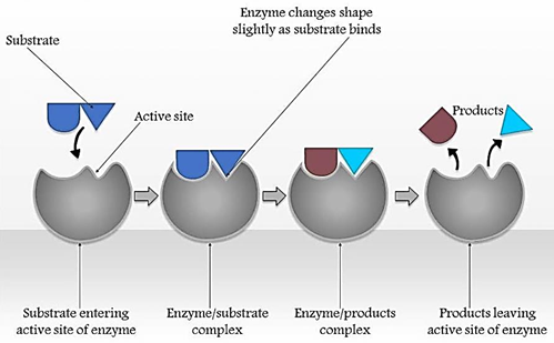
To complete the chemical reaction the energy barriers must be removed as these barriers hinder the spontaneous degradation of complex molecules such as nucleic acids and proteins. When the cell needed some metabolic change the breakdown of the complex molecules occurred, and these barriers were overcome by enzymes. Heat provided the additional activation energy, but the death of cells occurred due to a rise in the temperature. By the use of a catalyst, the level of activation energy was lowered. Enzymes react with the substrate and as a result, an intermediate complex was formed which was a transition state and it was quickly broken down to form the product and at last the enzyme was separated from the reaction [7].
Active sites were present on certain regions of an enzyme where substrate was attached. The active site was the groove which was developed due to the protein’s pattern of folding. The 3D structure of the enzyme having the electrical and chemical properties of amino acids and the presence of cofactor within the active site allowed the binding of the specific substrate with that active site. This determined the specificity of the enzyme for its activity and synthesis of enzyme which was under the influence of genetic control [40]. Some enzymes were only produced when they were required otherwise they were not produced in the cells and the enzymes were not uniformly found in the cell, most often they were present in the compartments in the nucleus, in subcellular structures, and in the cell membrane. There were certain factors such as hormones, neurosecretions, and many other chemicals that affect the internal surroundings of cells that were responsible for influencing the activity and synthesis of enzymes [7].
Figure 1. Mechanism of enzyme action, it describes how substrate reacts with the enzyme which results in the formation of enzyme-substrate complex finally this complex is converted into products and leaves the enzyme without any changes
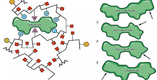
Figure 2. Binding of Active site with Substrate
Table 1. Table enlist some of the most industrially important enzymes showing multiple diagnostic application
|
Serum enzymes |
Diagnostic application |
|
Serum glutamate-oxaloacetate transaminase/ serum glutamate- pyruvate transaminase |
Viral hepatitis, Myocardial infarction |
|
Amylase |
Acute pancreatitis |
|
Ceruloplasmin |
Hepatolenticular degeneration |
|
Creatine kinase |
Muscle disorder, Myocardial infarction |
|
Lipase |
Acute pancreatitis |
|
Acid phosphatase |
Metastatic carcinoma of the prostate |
|
Alkaline phosphatase |
Bone disorder, obstructive liver diseases |
In the metabolic activity of animals, plants, humans, or microbes’ enzymes play an essential role and it had an extensive range of usage in microbial and diagnostical applications. The metabolic activity of enzymes has led to numerous metabolic disorders. From the studies, we came to know that a lot of diseases were associated with the components of the metabolism of enzymes, and they were used in clinical laboratories as a specific marker for the diagnosis of disease. There were some enzymes like Alanine transaminase, aspartate transaminase alkaline phosphatase, etc which had been used in clinical applications for the last 2 decades. In various diseases like myocardial infarction and jaundice etc, enzymes were used as preferable markers. Enzymes not only help in the diagnosis of a disease but also help in the understanding of prognosis and assessment of the response therapy [8].
There were few diagnostic applications of the enzymes
Certain enzymes were present only in specific tissues like lactase dehydrogenase which contain two different forms called isozymes and were present in the skeletal & heart muscle. These two forms of the enzyme differ only in the composition of amino acids. Lactase dehydrogenase was composed of four different types of subunits, but these enzymes were present in five different forms depending upon the source of the subunit [6]. An increase in the level of LDH may lead to tissue damage and if the level of LDH increases in the heart muscle then it may lead to a heart attack. Creatine kinase was another enzyme that was present in the heart, skeletal, and brain muscles and the appearance of brain type may cause brain tumors while heart type showed heart attack. The level of CK was higher as compared to LDH in the blood after the heart attack. The diagnosis of these enzymes could be made possible by proper monitoring because a mild heart attack was difficult to diagnose if it was not monitored properly [9]. The elevated level of isozyme might cause tissue damage. Acetylcholinesterase was another enzyme that played a role in the controlling of many nerve impulses. Many pesticides may affect this enzyme, so farmers make sure that they have not got any exposure to the toxin. Multiple enzymes have been used clinically for the diagnosis of multiple diseases. They might act as an important marker for the activity of enzymes in the liver, heart brain, etc. So, by the use of automated techniques, there was an ease to essay these enzymes, and that’s why doctors use the enzymes for disease diagnosis and treatment [10].
The rate of growth of bone was usually associated with alkaline phosphatase. The elevated level of serum alkaline phosphatase may indicate the active formation of bone or osteoblastic activity, as in the case of rheumatoid arthritis. The higher level of alkaline phosphatase may cause many pathological conditions like osteomalacia and rickets, etc [11].
In the male prostate gland, the level of AP was 100 times higher than other body tissues. In bone metastases, prostatic AP was expressed by prostate cancer cells. Abnormal expression of AP isoenzymes was valuable, especially in germ-cell tumors during the monitoring of cancers. The increased risk of hepatocellular carcinoma was associated with elevated alanine transaminase levels. In breast cancer, cathepsin D was useful in predicting women who were at risk for early recurrence in breast tissue. In human tumors, including breast and neck melanoma, etc, the enhanced expression of cysteine cathepsins has been demonstrated. In tumor progression, the presence of cyclooxygenase-2 played an essential role in early detection of tumorigenesis. The gastric cancer was closely related to the overexpression of glucose-6-phosphate dehydrogenase [9].
4.2. Enzymes Used in the Diagnosis of Cancer
A condition in which cells multiply and proliferate rapidly without any control is known as cancer. When the cells proliferate rapidly, it can be fatal, and the process is known as metastasis. In different body parts, cancer can occur, including the lungs, prostate, mouth, breast, brain, colon, or even in the blood. Certain enzymes were produced during the abnormal state of the body, and it was used as a biomarker of cancer prognosis. Multiple enzymes were used for the detection of cancer which are given below,
In humans, five different types of ACP were found in different parts/regions, namely prostatic, erythrocytic, macrophage, lysosomal, and osteoclastic. They have differences in their origin, molecular weight, sequence length, and resistance to tartrate and fluoride levels. Mainly, acid phosphatase was found in the prosthetic gland but was also found throughout the body. The ACP level was almost 100 times greater in the human male
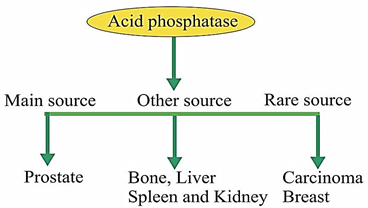
Figure 3. Sources for the production of acid phosphatases
prostate gland as compared to any other body tissue. For monitoring the progress of prostate cancer, the prostatic acid phosphatase was used because it was strongly expressed by prostate cancer cells. In semen, the acid phosphatases were present in very high concentrations, that’s why the rape victims were usually detected by the presence of acid phosphatase in the vaginal fluid [12].
Protein molecules were broken down into several polypeptide fragments, which can be digested easily by other lysosomal exo- and endopeptidases. This process was usually carried out by CD; it was a ubiquitous lysosomal aspartic protease. The CD was synthesized in the rough endoplasmic reticulum as preprocathepsin-D, and it was present in all cells, tissues, and organs except in lysosome-free RBC. CD-level was sometimes overexpressed in the epithelial breast cells, which led to the prognosis of breast cancer [13].
CCs were lysosomal protease enzymes that were involved in tumorigenic processes, i.e., invasion, apoptosis, and angiogenesis. 11 different types of CC enzymes were present in the human genome, and all of them show special types of expression that were responsible for particular physiological responses. At neutral pH, three types of CCs were stable, i.e., B, L, and H. If CCs were secreted out of their natural localization, then it would be harmful. In tumor conditions, the CC level was elevated, like in the case of the ovary, brain, head, uterine cancers, etc. Thus, it can be concluded that CCs were the most important biomarker in multiple inflammatory disorders such as inflammatory myopathies, periodontists in cancer, etc [14].
COX-2 was usually used for the conversion of arachidonic acid into prostaglandins H2, and during this process, multiple precursors were used, such as prostaglandins, thromboxanes, and prostacyclins. It was expressed in multiple types of processes, such as inflammatory reactions, apoptosis, carcinogenesis, immuno-suppression, cell proliferation, and invasiveness, which have been investigated through preclinical studies. COX-2 was also produced in response to mitogens, inflammatory mediators & RAS-mediated signaling cytokines. Nowadays, these enzymes have also been used as a biomarker for the progression of tumors, i.e., including stomach, breast, and urinary bladder carcinoma [15].
An analytical device called a biosensor was used to incorporate the biologically sensing materials that were closely related to the chemical and physical transducer. This device produced a continuous signal that was proportional to analyte concentration [16]. The first biosensor against the detection of glucose was developed in 1962. It was used as a transducer made of oxygen electrodes and was based on immobilized glucose oxidase. Then, the enzyme electrode concept became popular and applied in many other biosensors, which were based on enzymes and could detect a specific analyte. Thereafter, the clinical samples that contain urea can be detected from the potentiometric urea biosensor [17].
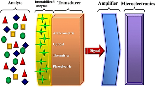
Figure 4. Enzyme-based biosensors; in this case, analytes are mixed with the immobilized enzyme and transducer and transmit the signal toward the amplifier and microelectronics
Table 2. Enzymatic Biosensors in Transducer Systems
|
Transducer |
Examples |
|
Electrochemical |
Clark electrode, medical electrode, ion selective electrode (ISE), field effect transistor (FET)- based devices |
|
Optical |
Photodiodes, waveguide systems, integrated optical devices |
|
Calorimetric |
Thermistor, thermopile |
|
Piezoelectric |
Quartz crystal microbalance (QCM), surface acoustic wave (SAW) devices |
|
Magnetic |
Bead-based devices |
For the biosensor construction, urease was immobilized in an ammonium ion electrode, which acted as a transducer. Moreover, for developing the biosensor, a thermistor was used as a transducer [18]. Optode was a term proposed by lubbers, and Opitz has a sensor made of fiber optic with an immobilized indicator that can be used for the measurement of oxygen and carbon dioxide. They have developed an optical biosensor, especially for alcohol, by immobilizing alcohol oxidase at the end of the fiber optic oxygen sensor [19].
A host of biosensors have been created to deliver patient health diagnostic information. The biosensors that can be used for diagnosis are mentioned below.
5.1. Biosensors for Blood Glucose
The deficiency of insulin in the body causes the disease known as diabetes. This disease was incurable and caused a decrease in the glucose level of blood or might also increase the glucose of blood. The reactions involving glucose metabolism were mediated by insulin, a hormone released from the pancreas [20]. A variety of medical disorders, such as cystic fibrosis, coeliac disease, heart disease, and tuberculosis, have been related to diabetes. Such conditions can lead to retinopathy, resulting in nephropathy, blindness causing the failure of the kidney, damage in the peripheral nerve, amputation, ulcers, cancer, and cardiovascular diseases. A silent epidemic was another name for diabetes. In the diabetic patient, the most studied analyte was glucose detection. Both in vitro and in vivo methods can be used for monitoring the glucose level. Many enzymes have been utilized for developing the glucose monitoring biosensors [21, 22].
5.1.1 Glucose Dehydrogenase-based Glucose Biosensors
For the 1st time, carbon paste and Erwinia sp. were used for developing the biosensor for glucose, and this biosensor was based on glucose dehydrogenase. Polylysine-albumin gel was used for the immobilization of the enzyme, and the paste of carbon was used as supporting material, binding material, and fumed silica was also used. The current of the anode from the biosensor on the glucose was observed as 0-200mv vs. Ag as a reference electrode. The emulsion of polyvinyl alcohol was used to coat the enzymatic layer of biosensors, which increased the biosensor linearity up to 3.0mM. At lower concentrations of glucose, a good linearity was shown by the developed biosensor because oxygen has been eliminated. 5-[2,5-Di-(thiophen-2-yl) -1H-pyrrol-1-yl] novel Electron-transfer Mediator The nicotinamide-oriented system of nicotinamide adenine dinucleotide-dependent dehydrogenase was synthesized -1,10- phenanthroline (III) chloride (PhenTPy). A disposable amperometric glucose biosensor was made with NAD-GDH on the carbon electrode screen print with FePhenTpy as a mediator for the transfer of electrons and its performance increase. Whereas at the same time, the performance of the sensor was further enhanced by adding the reduced graphene oxide. Then, in the experiment, Nafion containing lead acetate was coated on the surface of the sensor to avoid the interference effects of ascorbic acid, acetaminophen, uric acid, dopamine, monosaccharides, & caffeine. To check whether the glucose sensor was reliable or not, it was used to determine the glucose in human or artificial blood. In a recent study, a pencil graphite electrode was utilized for hybrid quantum dot (ZnS-CdS) deposition to construct a disposable biosensor. By using glutaraldehyde as a cross-linker, the immobilization of GDH was facilitated on the (GDH/ZnS-CdS/PGE) electrode. Cyclic voltammetry and photo-amperometric measurement have proved that when the surface of GDH/ZnS-CdS/PGE was uncovered by an optical source, GDH/ZnS-CdS/PGE might produce special signal photo-electrocatalytic activity to the NADH. There was a correlation between the glucose concentration and current produced by the enzymatic reaction in photo amperometric. Applied satisfactorily to the actual samples was the photoelectrochemical biosensor. The good results were shown by photo amperometric and amperometric transducer in monitoring the glucose. They show repeatability, sensitivity, and good selectivity [23].
5.1.2 Cellulose Dehydrogenase-based Glucose Biosensors
A cellulose dehydrogenase-based glucose biosensor was developed using the Corynascus thermophilus mutant, and the electrode deposition of gold nanoparticles was done to obtain the glassy carbon electrode. Then, monolayers of 4-amino thiophenol and 4-mercaptobenzoic acid were used to modify the electrode by cross link of these compounds. With 3.1±0.1 μA/mM cm2, a broad linear range was presented on the CDH/GA/4-APh, 4-MBA/AuNP/GCE platform, with an outstanding stability and strong glucose selectivity. This modified electrode was used for the detection of glucose up to 6.2 micrometers as the glucose biosensor. The glucose of humans can be detected from this biosensor from the Silva of humans as a sample. It also suggested the creation of non-invasive monitoring instruments for glucose [6].
5.2. Biosensors for Asparagine
L-asparaginase enzyme was used as a drug against tumors, lymphosarcoma, and acute lymphoblastic leukemia. This enzyme was not usually needed for the growth of normal cells. But during the growth of tumor cells, the amino acid L-asparagine was most important because tumor cells were deficient in asparagine synthetase, whichwas responsible for the production of L- asparaginases. L-asparaginase was required to stop the growth of tumor cells because it inhibited the supply of nutrients to tumor cells [24]. The most important application of L-asparaginase was that it was used as an injectable medicine into the blood, which can detect the level of L-asparagine in the blood of cancer-treated people to prevent recurrence. The nano level of L-asparagine in blood samples can be detected using the biosensor technology [6].
L-asparaginase is thermally stable and was usually isolated from Archaeoglobus fulgidus, and it can be expressed in Escherichia coli as a fusion protein. During the development of the biosensor, first of all, the L-asparaginase enzyme was immobilized on the surface of the ammonium selective electrode. The co-immobilization between the phenol red indicator and asparaginase was used to design an optical biosensor for the detection of asparagine [25].

Figure 5. L- Asparaginase detection
The maximum detection limit for asparagine was 10-10 M and 10-10 M for silicone gel and for calcium alginate pearls, and 10-9 M for other immobilization matrices, like nitrocellulose membrane. Moreover, asparagine levels in the leukemia blood and the blood of normal people were determined by a newly developed system. An oxacin 170 perchlorate (O17) ethyl cellulose membrane was used to produce a ratiometrically fluorescent L-asparagine biosensor, and the enzyme was imprisoned. The hydrolysis reaction, which was catalyzed by asparaginase, was the principle for developed biosensors, and this reaction can form ammonia. The ammonia generated by the O17-EC membrane interacted and altered the intensity of 625 nm of fluorescence. For asparagine, the detection limit of the developed biosensor was 0.074 ± 0.0023 mM, and the linear rand was 0.1-10 mM. The response, time, storage stability, and reversibility were also very good for sensing membranes. In the human blood sample, we can easily detect asparagine through developed methods [26].
Several billion years ago, there lived new antibiotics that were active against resistant bacteria. A wide range of antibiotics and drugs that occur naturally was found by them during that time. The resistance mechanism was developed by bacteria for their survival. There were two major characteristics which were very important of enzymes that distinguish them from other drugs. Firstly, there was great specificity and affinity between the binding of enzymes with their target. Secondly, enzymes, due to their catalytic activity, can change multiple molecules into the product of interest. Due to these two characteristics, enzymes were known as specific and potent drugs and can be used in therapeutics that other tiny molecules were unable to perform. These features of enzymes have made themselves used as drugs for treating a broad range of diseases or disorders [27].
Diagnostic importance was demonstrated in measuring serum levels of numerous enzymes. If the serum contained these enzymes, it meant that some cellular or tissue damage had happened, which led to the leaking of intracellular substances in the blood. Therefore, whenever a health care provider stated that he/she would test for liver enzymes, it was intended to determine the potential harm to the liver cells. The enzymes used for the assay include aspartate aminotransferase, AST, alanine transaminase ALT, lactate dehydrogenase LDH, gamma-glutamyl transpeptidase, and creatine kinase CK. Different clinical circumstances were used for the assay of other enzymes. In veterinary and humans, many diseases can be diagnosed using this enzyme [28]. The diseases can be diagnosed very quickly using these enzymes. The enzymes with different classes are mentioned below.
In 1920, alkaline phosphatase enzymes were discovered with the fact that they were divided to increase the diseases of the liver and bone, and they were known as very important for clinical purposes because phosphatase's earliest enzyme was found in serum. After that, instead of other enzymes, these were the subject of many publications. Many phosphate esters can be hydrolyzed using this alkaline phosphate. They were termed alkaline because they work in alkaline PH. The source for the ALP in the animal or human body includes the kidney, liver, and placenta bone. Many diseases like hepatobiliary and bone diseases were caused by it in humans. Bone or hepatic disease in cats and dogs can be diagnosed by the activity of total ALP serum. In liver illnesses of horses and ruminants, it was of limited utility because of the wide array of reference values to be compared with patients. The average value of serum ALP in goats was found to be 10-fold with no damage in the liver [29].
It was an organ-specific serum enzyme that can be used for therapeutic usage. They produce creatine phosphate by using ATP through reversible phosphorylation of creatine. Many body parts like the brain, heart, smooth muscles, and skeletal muscles contained creatine kinase, but their activity was very high in skeletal muscles. This enzyme can cause myocardial infarction in humans [30]. In some organisms, such as horses, dogs, cattle, and cats, the high level of this enzyme in cerebrospinal fluid may cause many disorders. We can find damage in the muscle through creatine kinase because it is very sensitive to that task. The clinical importance is only high elevations in serum activity [31].
Previously, this enzyme was also termed Glutamic Pyruvate Transaminase (GTP). It worked in the cytoplasm of the cell and produced glutamate and pyruvate through reversible transamination of 2-oxoglutarate and L-alanine. The liver, heart, and skeletal muscles contain the ALT. It specifically performed its activity in the liver of cats, rabbits, dogs, rats, and primates. It can sense any damage to the liver. It was less sensitive to diagnose tissue damage in pigs, cattle, horses, goats, or sheep. In toxicology investigations using tiny laboratory rodents as well as dogs, it was utilized to indicate hepatopathy [32].
7.4. Aspartate Aminotransferase
Previously, it was termed as Glutamic Oxaloacetic Transaminase. It produced glutamate and oxaloacetate through the transamination of 2-oxoglutarate and L-aspartate. Many organs, like the heart, kidney, liver, erythrocytes, and skeletal muscles contain AST [33]. In animals and humans, it may cause muscle, hepatic parenchymal, and myocardial diseases. The serum level of this enzyme in many tissues made it the best marker for soft tissues and organ-specific enzymes. AST was present in large amounts in RBCs, which when released in plasma before the hemolysis is indicated [34].
They were composed of 4 atoms of selenium and were known as metalloenzymes. They used peroxidase to oxidize the reduced glutathione and, as a result, produce oxidized glutathione and water. There existed a good correlation between the selenium concentration of organs and red blood cell GPx activity because these enzymes contained selenium in high concentration. Acid phosphate was another enzyme used for diagnosis purposes. Through the use of ACP, we can diagnose prostate carcinoma, and this enzyme was found in erythrocytes and the prostate. An enzyme that was involved in muscle disease was aldolase, and this enzyme was present in the heart and skeletal muscles [35]. Myocardial infarction was caused by hydroxybutyrate dehydrogenase, which was the cardiac version of lactate dehydrogenase. In humans and animals, enzyme assays were used to diagnose illnesses. This enzyme was also helpful in diagnosing plant diseases. For instance, there was an increase in the glucose six phosphate dehydrogenase instead of glucose phosphate isomerase during pathogenic or mechanical injury, resulting in the conversion of the pathway from glycolysis to pentose phosphate pathway [36].
Enzymes can be used in immunoassays as markers instead of using radioisotopes. In immunoassay, they can determine different hormones and proteins. The enzymes were used as markers in immunoassays because they were not hazardous and could be detected by more widely available procedures. For this purpose, enzymes were used whose assay procedure was sensitive and convenient. An example of enzyme immunoassay was the enzyme-multiplied immunoassay test and enzyme-linked immunosorbent assay. The detection of antibodies or antigens can be done using ELISA because it is a very sensitive assay. Through the use of ELISA, we can diagnose noninfectious diseases involving drugs, hormones, oncofetal proteins, serum components, or autoimmune diseases. Moreover, it has also found its application in diagnosing infectious diseases that were caused by viruses, Bactria, or parasites. ELISA uses the following enzymes: alkaline phosphate, Horseradish peroxidase, and B-galactosidase. In EMIT, malate dehydrogenase activity was assessed using a conventional thyroxin detection enzyme-balanced immunoassay approach [30] [37].
Many medical problems can be treated by giving therapy of the enzymes as drugs. Streptococcus was used in the preparation of a mixture of enzymes called streptokinase. It was beneficial in removing clots of blood. Streptokinase performed its action by activating the plasminogen that was found in plasma. Plasmin was the active form of this enzyme called serine proteases [38]. It was similar to trypsin and acted to cleave the fibrin into the soluble parts. Asparaginase was also a therapeutic enzyme. For the treatment of leukemia, asparaginase therapy was used. There was a need for nutrition for the asparagine to scavenge the tumor cell from the plasma of the host. The plasma level of asparagine was significantly reduced by the administration of asparaginase; hence, tumor viability was reduced [39].
The other application of therapeutic enzymes is that they can be used as an enzyme replacement in the enzyme deficient person. Urokinase was the enzyme, also called u-plasminogen activator, which was isolated from the urine of humans and was injected into a patient who was suffering from pulmonary embolism. An active plasmin was produced by these enzymes when they stimulated the cascade system. Plasmin has proteolytic activity, and it helps in digesting fibrin. We can also use these enzymes to stop the cancer cells from growing by stopping the supply of nutrients to these cells. For instance, different types of leukemia can be treated using L-asparaginase. The L-asparagine was the requirement of tumor cells, not the normal cells. The use of immobilized enzymes as artificial kidney machine ingredients used to eliminate urea and other waste materials from the body was another example of the therapeutic application of enzymes. Immobilized urease can be used in converting urea into NH4+ and CO2 when the blood from the body is entered into the machine by dialysis. Then glutamate dehydrogenase can entrap the toxic NH4+ on ion exchange resins and is then absorbed into glutamate by the action of this enzyme. Then, the coenzyme is recycled through alcohol dehydrogenase before the fluid is given back to the blood [40].
Enzymes and enzyme-based biosensors play an important role in the diagnosis of various disorders due to their rapid response, specificity, sensitivity, and portability. The high specificity of the enzymes made them a good candidate for medical diagnosis. Most of the research that was carried out on the enzyme used in the clinical diagnosis was performed on the laboratory scale. Multiple enzymes were used in the diagnosis of cancer, such as acid phosphatases, cathepsin D, cysteine cathepsins, and cyclooxygenase-2. Enzymes can be used as biosensors that can detect levels of glucose and L- asparagine. Similarly, enzymes have versatile applications in pathology, immunoassay, drug/antibiotics, and therapeutic agents. Glucose biosensor was the commercially available biosensor used for glucose level detection in the blood samples. With the advancement in the use of enzyme technology, it will become an important tool in the diagnosis and detection of diseases like epilepsy, cancer, heart failure, etc.