Study of Copper Bioremediation by Planktonic Cells and Biofilms of Bacteria Isolated from Indigenous Environment
Sanam Asma1, Hafiz Zeshan Wadood1*
|
KEYWORDS Atomic absorption spectrophotometer, Biofilms, Bioremediation, Minimum inhibitory concentration |
AbstractThis research aimed to isolate copper-resistant bacteria from industrial effluents for potential bioremediation in both planktonic and biofilm growth modes. Out of ten isolates from industrial effluents, four bacterial strains (S1A, S2C, SSA, and S1C) were selected based on their minimum inhibitory concentration (MIC) and biofilm-forming capabilities. These bacteria demonstrated strong biofilm formation abilities in both the absence and presence of copper (Cu) stress, with MIC values of 850 µg/ml for S1A, SSA, and S1C, and 750 µg/ml for S2C. Physiological characterization revealed that these isolates exhibited optimal growth at pH 7 and 37°C. Biochemical characterization indicated the similarity of these copper-resistant bacteria with the genera Staphylococcus (S1C), Bacillus (SSA), Corynebacterium (S1A), and Enterobacter (S2C). The copper removal efficiency of these isolates was assessed in both planktonic and biofilm growth modes using atomic absorption spectroscopy. In planktonic growth, all isolates showed copper removal efficiencies of 81.4% (S1A), 81% (SSA), 83.5% (S2C), and 82.3% (S1C) after 24 hours, and 84% (S1A), 83.4% (SSA), 85.3% (S2C), and 84.2% (S1C) after 48 hours. Notably, in planktonic growth, S2C (Staphylococcus) exhibited the highest removal efficiency, with 83.5% and 85.3% after 24 and 48 hours, respectively. In the biofilm growth mode, copper removal efficiencies were 84.2% (S1A), 82.7% (SSA), 81.9% (S2C), and 84% (S1C) after 24 hours, and 86.7% (S1A), 86.1% (SSA), 85.6% (S2C), and 86.2% (S1C) after 48 hours. Notably, S1A (Corynebacterium) displayed the highest copper removal efficiency, with 84.2% and 86.7% after 24- and 48-hour incubation in biofilm growth modes. |
The continuous discharge of heavy metal-laden waste into the environment can lead to significant medical and financial problems.[1] Heavy metals are known to damage cell membranes and disrupt enzyme activity within cells.[2] Wastewater from landfills often contains toxic heavy metals, and using this wastewater for irrigation can result in heavy metal contamination of food crops and farmland. Various toxic heavy metals, including lead, zinc, copper, chromium, cadmium, nickel, and arsenic, can be present in wastewater.
Copper's persistence in the environment prevents its breakdown, potentially leading to its accumulation in plants and animals residing in copper-rich soils. In such areas, only a limited variety of plants can thrive, which reduces plant diversity near factories emitting copper. The impact of copper on plant life poses a significant threat to agricultural production.
Copper can disrupt soil activity by adversely affecting microorganisms and earthworms, leading to a considerable slowdown in the decomposition of organic matter. In soil, copper plays a role in organic matter decomposition and pest control. The concentration of copper increases in soil due to metal-producing industries, which can harm plants. Excessive copper is detrimental to beneficial soil microbes that support plant growth.[3] It's important to note that copper poisoning primarily affects living organisms, even at relatively low concentrations, potentially causing liver and kidney damage and even fatal outcomes. Exposure to industrial copper emissions, dust, or mists can lead to metal fume fever, accompanied by atrophic changes in nasal mucous membranes. Chronic copper poisoning can result in Wilson's disease, characterized by hepatic cirrhosis, brain damage, demyelination, renal disease, and copper deposition in the cornea. Heavy metals discharged into the environment are harmful to humans, animals, aquatic organisms, and plants, increasing pollution of both animals and plants.[4]
Copper is an essential trace element for plants; at low concentrations, it plays a catalytic and structural role in plant development and growth. High copper levels in the environment can adversely affect the photosynthetic process in plants.[5] Copper, as a metallic element, becomes toxic to bacteria at high concentrations. Through the discharges of copper from factories and mining sites, copper can enter the environment and impact plants, animals, and humans.[6]
Understanding copper resistance mechanisms in bacterial species can be a valuable tool for the remediation of copper from the environment, a process known as bioremediation.[7]
Bioremediation is the process of using microorganisms and biological mechanisms within plants to remove harmful pollutants and restore the environment to its original state, effectively eliminating pollutants from the environment. The bioremediation process employs various microbes for the degradation of exogenous compounds, heavy metals, herbicides, crude oil, petroleum products, aromatic hydrocarbons, volatile organic compounds, pesticides, radionuclides, kerosene, and explosives in the environment.[8] Bioremediation can be categorized into two types based on the location of pollutant treatment: in-situ and ex-situ bioremediation.[9] In in-situ bioremediation, contaminated samples are processed underground, while in ex-situ bioremediation, contaminated samples are processed in cells and bioreactors.
Microbial strains that efficiently degrade heavy metal pollutants are used in bioremediation, making it a crucial method for removing heavy metals from contaminated sites.[10] Bioremediation is cost-effective, easy to manage, and does not lead to secondary pollution. Bacterial biofilms have the ability to survive and persist within indigenous populations, even in harsh environments. Biofilms protect bacteria for extended periods under normal environmental conditions due to their coverage with extracellular polymeric substances (EPS). These substances, which include polysaccharides, proteins, lipids, nucleic acids, and water, play a vital role in producing extracellular polymeric substances (EPS).[11]The biofilm matrix provides greater resistance against shear stress, UV damage, antimicrobial agents, and high concentrations of toxic chemicals and heavy metal pollutants compared to planktonic cells.[12] Biofilms are particularly useful for treating heavy metals such as copper, cadmium, uranium, and chromium, which are harmful to the environment and serve as toxic pollutants.
The main objective of this research project was to isolate and characterize copper-resistant bacteria from the local environment. The biofilm-forming abilities of these copper-resistant bacteria were assessed through qualitative and quantitative analyses of biofilm formation. The Minimum Inhibitory Concentration (MIC) of all copper-resistant bacterial cells against different copper concentrations was determined. The copper absorption capabilities of biofilm-based bacterial cells and planktonic-based bacterial cells were measured using atomic absorption spectrophotometry.
Sample Collection
Two types of samples, namely wastewater and soil samples, were collected from the industrial area of Lahore, Punjab. Wastewater samples were obtained from the drains of the Rado textile industry and Lahore dying industry, both situated in the Quaid-e-Azam Industrial Estate Kot Lakhpat, Lahore, Punjab. Wastewater samples were collected in sterile bottles, while soil samples were collected from an area adjacent to the drains in sterile plastic bags. Physicochemical parameters, such as pH and temperature, were measured at the time of sample collection.
For the isolation of copper-resistant bacteria, wastewater and soil samples were serially diluted and plated on nutrient agar plates supplemented with copper sulfate stress (100 µg/ml). These plates were then incubated at 37°C for 24 hours. After incubation, distinct colonies with varying morphologies were selected for further purification.
Bacterial isolates cultivated on copper-supplemented media were characterized based on their morphological features, including colony morphology and cell morphology. Colony morphology, including size, color, shape, elevation, and margins, was examined using pure colonies of bacterial isolates on nutrient agar supplemented with copper stress. For the assessment of cellular morphology, including cell shape and arrangement, the copper-resistant bacterial isolates were subjected to Gram staining.
The qualitative assessment of biofilm formation by bacterial isolates was conducted following the procedure outlined by ÇAM APDSJIAFM, 2022.[13]
In brief, bacterial cultures were grown by inoculating 100 μl of overnight bacterial cultures (OD600nm = 0.1) into test tubes containing 10 ml of L-broth, with one set supplemented with 100 μg/ml of Cu+2 stress, and another set without Cu+2 stress. Negative controls (media without any inoculum) were included. The tubes were incubated at 37°C without shaking for 72 hours. Afterward, the liquid medium was removed, and the tubes were stained with a 0.1% crystal violet aqueous solution for 20 minutes to visualize the bacterial biofilm. Excess stain was removed, and the test tubes were rinsed with sterile deionized water. Biofilm formation (identified as a purple ring) was observed after the test tubes were air-dried.
A quantitative assay for biofilm formation of bacterial isolates was performed in sterile polystyrene 96-well flat-bottom microtiter plates in triplicates, following the protocol described by Rao TS, 2022.[14] Autoclaved L-broth without Cu+2 stress was added to two rows of microtiter plates (180 µl/well), and L-broth with Cu+2 stress was added to the other two rows. A 20 μl inoculum of bacterial culture (OD600nm= 0.1) was added to all wells, except control wells. Control rows included one with L-broth without +2 stress and bacterial culture and one with L-broth with Cu+2 stress but without bacterial culture. The microtiter plates were sealed and incubated at 37°C for 72 hours without agitation. After incubation, the medium in each well was carefully removed, ensuring that the biofilm was not disturbed. The biofilm was fixed with 99% methanol, the plates were washed with distilled water, and then air-dried. Subsequently, a 0.1% aqueous crystal violet solution was added to each well and left for 20 minutes. Excess crystal violet was removed, and the plates were washed with distilled water and air-dried. Cell-bound crystal violet was dissolved in 33% glacial acetic acid, and the biofilm's growth was measured by its optical density at 570 nm using an ELISA microtiter plate reader (BIO-RAD iMark Tm).
A broth dilution method was used to determine the MIC of copper for the bacterial isolates, following the procedure described by Bezza FA et al,. 2020.[15]
LB broth was prepared in test tubes (10 ml per tube) and supplemented with varying concentrations of Cu+2 (ranging from 100 µg/ml to 1000 µg/ml) with a 50 µg/ml gap between two concentrations. Each tube was inoculated with 100 μl of culture per tube using a standardized (OD600nm= 0.1) overnight bacterial culture. A negative control without inoculum was included for each metal concentration. The inoculated tubes were incubated at 37°C for 24 hours at 100 rpm, and MIC was defined as the minimum copper concentration that inhibited visible bacterial growth.
The influence of temperature and pH on the growth of bacterial isolates was assessed following the procedure outlined by Gopi K et al,. 2022. Biochemical characterization of the bacterial isolates was carried out in accordance with the protocols described.[16]
Estimation of Copper Biosorption by Planktonic Bacterial Isolates
The estimation of copper biosorption was conducted following the procedure described by Tahmourespour A, 2021.[17]
For planktonic cells, L-broth (250 ml per flask) was prepared, autoclaved, and supplemented with 100 µg/ml Cu+2 stress. The flasks were then inoculated with a 1% inoculum per flask using a standardized (OD600nm = 0.1) 24-hour-old incubated bacterial culture. Negative controls were also included in this experiment. Two sets of flasks were inoculated using the same scheme, one for 24 hours of incubation and one for 48 hours. The flasks were incubated at 100 rpm and 37°C for their respective incubation times. After the designated incubation time, the cultures were transferred to sterile centrifuge tubes and centrifuged at 5000 rpm for 20 minutes. The supernatants were stored at refrigerated temperature until processed for copper estimation by atomic absorption spectroscopy.
Estimation of Copper Biosorption by Biofilms of Bacterial Isolates
For the formation of biofilms, L-broth medium with immersed glass slides was prepared and autoclaved. An inoculum (1%) of standardized bacterial cultures (OD600nm = 0.1) was added to the respective flasks designated for each bacterial isolate, and the flasks were incubated at 37°C for 48 hours without agitation. After 48 hours of incubation, biofilms formed on the glass slides were used as the inoculum in the flasks designated for estimating copper biosorption by biofilms of bacterial isolates. For biofilms, L-broth (250 ml per flask) was prepared, autoclaved, and supplemented with 100 µg/ml Cu+2 stress. The inoculum of biofilms (glass slides with formed biofilms) was added to the L-broth medium supplemented with 100 µg/ml copper stress for each bacterial isolate. Two sets of flasks were inoculated using the same scheme, one for 24 hours of incubation and one for 48 hours. These flasks were incubated at 37°C without agitation for their respective incubation time periods (24 and 48 hours). After the completion of the respective incubation times, cultures were transferred to sterile centrifuge tubes and centrifuged at 5000 rpm for 20 minutes. The supernatants were stored at refrigerated temperature until processed for copper estimation by atomic absorption spectroscopy.
Each supernatant was used to estimate copper concentration using an atomic absorption spectrophotometer (λ=249.2nm).
RESULTS
Certain physicochemical parameters of the wastewater, specifically temperature and pH, were recorded at the sampling site. It was observed that the temperature of the wastewater samples ranged from 36 to 46°C, and the pH values fell within the range of 8 to 9.
Isolation and Purification of Copper-resistant Bacteria
A total of ten bacterial isolates were chosen, with nine originating from wastewater samples and one from a soil sample, based on variations in the morphology of bacterial colonies. These selected bacterial isolates were subjected to multiple rounds of streaking on nutrient agar plates supplemented with copper (100 μg/ml) to achieve purification. The morphological characteristics of the isolated bacterial colonies are provided in Table 1.
Determination of Biofilm Formation of Bacterial Isolates
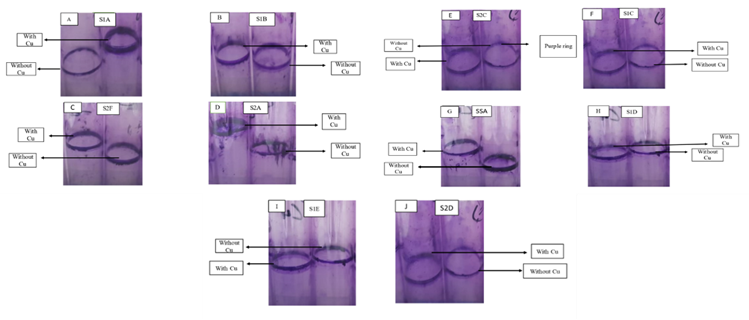
The biofilm-forming ability of the bacterial isolates was qualitatively assessed using a ring test, both in the presence and absence of copper stress. The results revealed that all these bacterial isolates were capable of forming biofilms, with varying degrees of biofilm formation. Biofilm formation was identified by the presence of a dark purple ring on the surface of the test tube (Figure 1).
Table 1: Morphological characteristics of the isolated copper resistant bacterial colonies
|
Bacterial isolates |
Size |
Color |
Form |
Margins |
Elevation |
Gram staining |
Arrangement and shape of cells |
|
S1A |
moderate |
Brownish |
circular |
entire |
convex |
positive |
irregular rods |
|
S1B |
moderate |
Brownish |
circular |
undulate |
umbonate |
positive |
irregular rods |
|
S2C |
pinpoint |
Yellow |
circular |
entire |
raised |
negative |
irregular rods |
|
SSA |
pinpoint |
off-white |
circular |
entire |
flat |
positive |
irregular rods |
|
S1C |
small |
Yellow |
circular |
entire |
convex |
positive |
clusters cocci |
|
S2D |
small |
Yellow |
circular |
entire |
raised |
negative |
clusters cocci |
|
S2F |
small |
off-white |
circular |
undulate |
convex |
negative |
irregular rods |
|
S1D |
small |
White |
circular |
entire |
flat |
positive |
irregular rods |
|
S1E |
small |
White |
circular |
undulate |
umbonate |
positive |
irregular rods |
|
S2A |
large |
Transparent |
rhizoid |
lobate |
umbonate |
positive |
irregular rods |
Figure 1: Qualitative assay of biofilm formation by ring test
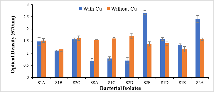
Figure 2: Quantitative analysis of biofilm formation in the presence and absence of copper stress
Quantitative Analysis of Biofilm Formation
The copper-resistant bacterial isolates underwent quantitative analysis to assess their biofilm formation abilities, and the results of the quantitative assay were consistent with the qualitative assay. The graph (Figure 2) below illustrates that all ten bacterial isolates (S1A, S1B, S2C, S1C, SSA, S1D, S2F, S2D, S1E, S2A, and S2F) are capable of forming biofilms.
The results of the Minimum Inhibitory Concentration (MIC) are presented in Figure 3. Among the bacterial isolates, SSA, S1C, and S1A exhibited MIC values of 850 µg/ml, while S2C had a MIC value of 750 µg/ml. Additionally, S2D, S1B, S2F, S1E, S1D, and S2A had MIC values of 650 µg/ml, 550 µg/ml, 450 µg/ml, 350 µg/ml, and 250 µg/ml, respectively (Figure 3). Notably, bacterial isolates SSA, S2C, S1A, S1C, and S2D demonstrated a high MIC against copper stress.
The results depicting the influence of pH and temperature on the growth of the selected bacterial isolates are presented in Figure 4 and Figure 5, respectively. Among these isolates, SSA, S2C, and S1C displayed an optimal pH of 7, while S1A exhibited an optimal pH value of 9. Notably, all bacterial isolates, including S1A, SSA, S2C, and S1C, demonstrated their maximum growth at 37°C, signifying that 37°C was the optimum temperature for these bacterial isolates.
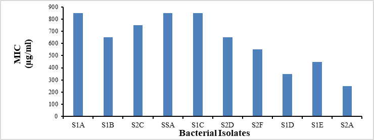
Figure 3: Minimum inhibitory concentration (MIC) for Copper-resistant bacterial isolates
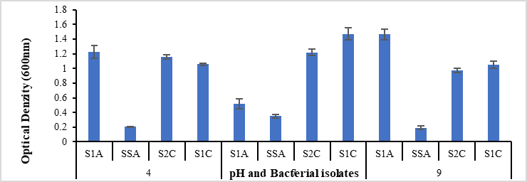
Figure 4: Effect of pH on the growth of copper-resistant bacterial isolates
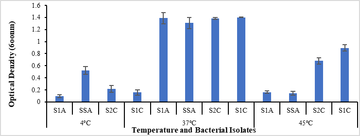
Figure 5: Effect of temperature on the growth of copper-resistant bacterial isolates
Table 2: Biochemical characteristics of the selected copper resistant bacterial colonies
|
Bacterial isolates |
S1A |
S1C |
S2C |
SSA |
|
Catalase test |
+ |
+ |
+ |
+ |
|
Oxidase test |
- |
- |
- |
- |
|
Indole test |
- |
- |
- |
- |
|
MR test |
+ |
+ |
- |
+ |
|
VP test |
- |
- |
+ |
- |
|
Citrate test |
- |
- |
+ |
+ |
The results of biochemical tests of copper resistant bacterial isolates is given in Table 2.
Based on the biochemical characterization of copper-resistant bacteria, the bacterial isolates exhibited a close resemblance to other bacterial genera. Specifically, S1C displayed a strong resemblance to the Staphylococcus genus, S1A exhibited a close similarity to the Corynebacterium genus, S2C showed a resemblance to Enterobacter, and SSA demonstrated a likeness to the Bacillus genus.
Copper Removal by Bacterial Isolates in Planktonic and Biofilm Modes of Growth
The copper removal potential of bacterial
isolates were assessed in both planktonic and biofilm growth modes after 24 and 48 hours of incubation, using atomic absorption spectrophotometry. The graph below illustrates that in their planktonic growth mode after 24 hours, bacterial isolates S1A, SSA, S2C, and S1C removed 81.4 µg/ml, 81 µg/ml, 83.5 µg/ml, and 82.3 µg/ml of Cu+2 from the culture media, respectively. After 48 hours under the same conditions, these isolates removed 84 µg/ml, 83.4 µg/ml, 85.3 µg/ml, and 84.2 µg/ml of Cu+2, respectively.
In the biofilm growth mode after 24 hours, the bacterial isolates S1A, SSA, S2C, and S1C removed 84.2 µg/ml, 82.7 µg/ml, 81.9 µg/ml, and 84 µg/ml of Cu+2, respectively. After 48 hours, these isolates removed 86.7 µg/ml, 86.1 µg/ml, 85.6 µg/ml, and 86.2 µg/ml of Cu+2 (Figure 6).
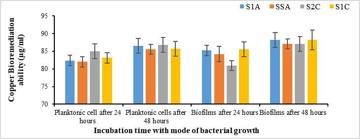
Figure 6: Copper removal efficiency of copper resistant bacteria in planktonic and biofilm mode of growth
The concurrent pollution of organic pollutants and metals is a significant global concern, as these pollutants pose threats to living organisms.[18]While copper is considered an essential trace element, elevated concentrations can have detrimental effects on living beings, causing harm. Various conventional methods, both physical and chemical, have been employed to remove excessive copper; however, bioremediation is considered a more effective, cost-efficient, and eco-friendly approach for tackling high copper concentrations.[19]
In the present research study, samples of two types, wastewater and soil, were collected from the Quaid-e-Azam Industrial Estate in Lahore, Punjab. A total of ten copper-resistant bacteria were screened and purified from these samples using a medium supplemented with 100 µg/ml copper stress. Nine copper-resistant bacteria (S1A, S1B, S2C, S1D, S2F, S1C, S1E, S2A, and S2D) were isolated from wastewater samples, while one copper-resistant bacterium (SSA) was isolated from the soil sample. Previous studies have also isolated copper-resistant bacteria from industrial wastewater and characterized the isolates for their copper resistance. These copper-resistant bacterial isolates were morphologically characterized (Table 2), including observations of colony morphology such as size, color, form, margin, and elevation. In terms of cell morphology, Gram staining was performed to observe cell shape and arrangement. The results of Gram staining indicated that six bacterial isolates (S1A, S1B, SSA, S1E, S1D, S2A) were Gram-positive rods, two bacterial isolates (S2C and S2F) were Gram-negative rods, one bacterial isolate (S1C) was Gram-positive cocci, and one bacterial isolate (S2D) was Gram-negative cocci.
The minimum inhibitory concentration (MIC) of copper-resistant bacterial isolates against copper was determined, and all the bacterial isolates exhibited varying MIC values (Figure 3). Four bacterial isolates, S1A, SSA, S1C, exhibited an MIC of 850 µg/ml, while S2C had an MIC value of 750 µg/ml. These were the highest MIC values among the bacterial isolates. Three bacterial isolates, S1B and S2F, showed MIC values of 650 µg/ml, and S2D exhibited an MIC value of 550 µg/ml. The remaining three bacterial isolates, S1D, S1E, S2A, had the lowest MIC values of 450 µg/ml, 350 µg/ml, and 250 µg/ml, respectively. Other researchers have also isolated copper-resistant bacteria and determined their MIC against copper stress. For instance, Enterobacter and Pseudomonas showed high MIC values of 2mM and 1 mM, respectively.20 Klebsiella exhibited the maximum MIC value of 6 mM.7 Enterobacter and Escherichia coli displayed the highest MIC value of 280 µg/ml.[21]
In the formation of biofilms, bacterial cells release extracellular polymers (EPS) that bind cells and extracellular materials together, resulting in high cell density, which accelerates bioremediation.[22] In this research, the biofilm-forming ability of all isolated copper-resistant bacterial cells was assessed both qualitatively and quantitatively. This analysis revealed that all bacterial isolates had the ability to form biofilms. Some isolates, such as S2F and S2A, were found to form firm and rigid biofilms under copper stress, but weaker biofilms without copper stress. Other bacterial cells, like SSA, S1C, and S2D, formed strong and firm biofilms without copper stress, while producing thinner biofilms under copper stress. In contrast, isolates such as S1A, S1B, S2C, S1E, and S1D formed strong and rigid biofilm rings both with and without copper stress.
Based on their MIC values and biofilm-forming abilities, four bacterial isolates (S1A, SSA, S2C, and S1C) were selected for further investigation. These selected bacterial cells underwent physiological and biochemical characterization. Biochemical characterization revealed that all four selected bacterial isolates (SSA, S1A, S1C, and S2C) belonged to four different genera, namely Corynebacterium (S1A), Bacillus (SSA), Staphylococcus (S1C), and Enterobacter (S2A).
In the current research, all four bacterial isolates were optimized for different temperatures and pH levels. Each of the four isolates exhibited the most growth at 37°C, making 37°C the optimal temperature for all selected bacterial isolates. Furthermore, the isolates were allowed to grow at different pH levels, including 4, 7, and 9, to determine the optimum pH for copper-resistant bacteria. All four isolates exhibited the highest growth at a pH of 7, indicating that the optimum pH for all selected bacterial isolates was 7.
Additionally, the copper absorption ability of the four selected bacterial isolates was assessed and statistically analyzed. The copper removal efficiency of these isolates was determined in both planktonic and biofilm growth modes using atomic absorption spectrophotometry. The results indicated that after 24 hours, bacterial isolates S1A (Corynebacterium), SSA (Bacillus), S2C (Enterobacter), and S1C (Staphylococcus) removed 81.4%, 81%, 83.5%, and 82.3% of Cu, respectively, in planktonic form. After 48 hours under the same conditions, these isolates removed 84%, 83.4%, 85.3%, and 84.2% of Cu, respectively, from the culture media in their planktonic growth mode. These findings suggest that overall, the bacterial isolates removed more copper after 48 hours of incubation compared to 24 hours. In the case of individual bacterial isolates, S2C (Enterobacter) exhibited the highest copper removal efficiency, removing 83.5% of copper from the media after 24 hours, which remained consistent after 48 hours. In contrast, isolate SSA (Bacillus) showed the lowest copper removal efficiency, at 81% and 83.4% after 24- and 48-hour incubations in planktonic growth mode. Similar studies have also been conducted by other researchers, who employed Klebsiella pneumoniae and Acinetobacter calcoaceticus for copper removal, with these bacteria demonstrating high copper removal efficiencies of 36.7% and 48.15%, respectively.[7]
In the biofilm growth mode, bacterial isolates S1A (Corynebacterium), SSA (Bacillus), S2C (Enterobacter), and S1C (Staphylococcus) removed 84.2%, 82.7%, 81.9%, and 84% Cu+2, respectively, from the media after 24 hours of incubation. The same bacterial isolates, after 48 hours of incubation, removed 86.7%, 86.1%, 85.6%, and 86.2% copper, respectively, from the culture media. For individual bacterial isolates, S1A (Corynebacterium) removed the highest amount of copper (84.2%) from the media after 24 hours, and this rate increased to 86.7% after 48 hours of incubation in the biofilm growth mode. Conversely, S2C (Enterobacter) exhibited the lowest copper removal efficiency, at 81.9% and 85.6%, after both 24- and 48-hour incubations in the biofilm growth mode. Numerous researchers have also investigated the copper removal efficiency in the biofilm mode of bacterial growth. Staphylococcus spp. and Pseudomonas demonstrated high copper removal efficiencies of 65% and 80%, respectively.[17] The selected isolates, S1A (Corynebacterium), SSA (Bacillus), S2C (Staphylococcus), and S1C (Enterobacter), displayed maximum copper removal ability, with rates of 86.7%, 86.1%, 85.6%, and 86.2%, respectively, after 48 hours in the biofilm growth mode, compared to the planktonic growth mode.
The primary objective of this study was to assess the effectiveness of copper-resistant bacteria in remediating copper, both in their planktonic and biofilm growth modes. The ability of bacterial isolates to remediate copper was determined using atomic absorption spectroscopy. It was observed that after 48 hours, bacterial isolates exhibited greater copper remediation compared to a 24-hour incubation period. Furthermore, when comparing the planktonic and biofilm growth modes, all four bacterial isolates (S1A - Corynebacterium, SSA - Bacillus, S2C - Staphylococcus, and S1C - Enterobacter) showed their highest copper remediation capabilities in the biofilm growth mode. Specifically, they were able to remediate 86.7 µg/ml, 86.1 µg/ml, 85.6 µg/ml, and 86.2 µg/ml of copper, respectively, in their biofilm modes, surpassing their planktonic growth modes after both 24 and 48 hours of incubation.
For future research, it is recommended to delve into the genetic and metabolic mechanisms underlying these remediation processes, assess potential ecological impacts, explore biotechnological applications, and conduct comparative studies with other remediation methods. Additional recommendations include optimizing growth conditions, investigating the potential of microbial consortia, developing efficient bioreactors, raising public awareness, addressing regulatory and safety considerations, and establishing long-term monitoring practices. These actions will enable us to fully harness the potential of copper-resistant bacteria in bioremediation and contribute to sustainable solutions for copper pollution.
1Department of Biology, Lahore Garrison University, Lahore, Pakistan
*Correspondence: Shan_wadood@yahoo.com