Characterization and Prevalence of Antibiotic Resistance Staphylococcus aureus in Street Food: A Public Health Concern
Arooba Siddique1 | Saher Mahmood1* | Sadia Tahir1 | Izna Tariq1 | Chaudhry Ahmed Shabbir2 | Yasir Arfat3
|
1Department of Microbiology and Molecular Genetics, The Women University Multan, Pakistan 2Department of Molecular Pathology, Osaka Metropolitan University 3Department of Microbiology, Government College University Faisalabad, Pakistan *Correspondence Saher Mahmood Email: saher.9006@wum.edu.pk |
|
ABSTRACT Food samples containing Staphylococcus aureus pose a serious health risk. This study aimed to examine the prevalence of S. aureus strains in various food samples sourced from Mailsi and Multan. Many food samples including yogurt, bakery products, and raw and cooked food were examined for S. aureus. The isolated strains were confirmed through Mannitol agar fermentation, catalase, coagulase, and urease tests. Hemolysis on blood agar and biofilm formation were also assessed to determine toxin production. Antibiotic sensitivity testing was conducted using the Kirby Bauer method on MH agar, and multiple antibiotics were tested. Out of all samples, a total of 50 S. aureus strains were obtained, mainly from milk and milk-based products. Yellow colonies on mannitol salt agar confirmed S. aureus presence, with all isolates testing positive for coagulase, catalase, and urease. The presence of hemolysins: beta, gamma, and alpha were revealed by hemolysis assays. Biofilm assay results showed variation among the strains, with some categorized as strong, moderate, or weak biofilm formers. Regarding antibiotic sensitivity, most strains exhibited multidrug resistance, particularly against certain antibiotics. Vancomycin showed varying susceptibility patterns, some strains showed susceptibility and intermediate resistance, whereas only milk samples showed resistance. This study emphasizes the prevalence of MDR S. aureus strains in food samples. The study underscores the significance of antimicrobial stewardship programs and stringent food safety measures in preventing the spread of antibiotic-resistant strains and reducing foodborne illnesses. Further research is needed to explore the mechanisms behind antibiotic resistance and toxin production in S. aureus strains from food samples. |
|
Keywords: Staphylococcus aureus Food, Hemolysis assays, biofilm, food safety, Antibiotic, Public health |
A dreadful pathogenic microorganism, namely Staphylococcus aureus, lives in the skin and nasal membranes and is responsible for many hospital-acquired and community infections [1]. Staphylococcus aureus is responsible for various severe diseases accompanied by clinical presentation ranging from small skin infections to life-threatening infections [2]. In community settings and health institutions, S. aureus pathogenicity has become a great threat with the emergence of methicillin-resistant S. aureus [3]. The colonies of methicillin-resistant S. aureus have been identified in food such as milk products and raw milk [4]. In humans, the principal ecological niche of this bacterium is anterior nares, where it colonizes. S. aureus colonies can only be stored at 4 degrees Celsius [5]. This bacterium is characterized by high virulence and flexibility, allowing it to cause infection, invasion, persistence, and replication in various human tissues like skin, bone, or visceral organs [6]. Staphylococcus aureus produces catalase, which breaks down hydrogen peroxide, thus aiding its survival in the host by protecting it from the host’s immune system. Unlike other Staphylococcus species, the coagulase enzyme is produced by all isolates of S. aureus which is a virulence factor and aids in recognizing this bacteria [7]. Upregulated expression of various toxins like gamma toxin, by the Agr gene, helps S. aureus in adhesion and biofilm formation [8]. Unhygienic practices in food processing industries may result in food contaminated with pathogens that impose a serious health hazard for consumers. In many food sectors, biofilms are a severe problem. In the food industry, S. aureus can thrive in hostile environments due to its ability to form biofilms [9]. The milking equipment contaminated with biofilm is a great source of S. aureus [10]. It can be transferred to food from air, food contact surfaces, and dust. It can grow at low water activity and has also been found in salted food. An intoxication that is reported by eating food possessing one or more preformed toxins is referred to as Staphylococcal food poisoning [11]. Symptoms of acute gastroenteritis arise in humans when exoproteins, namely Staphylococcal enterotoxins (SE), are ingested. Enterotoxins are low molecular weight proteins. SEs are pyrogenic toxin antigens characterized by high stability, showing resistance to more proteolytic enzymes like trypsin or pepsin and thus retaining their activity in the digestive tract of humans [12]. The test food may be negative for S. aureus but positive for S. aureus enterotoxins, as enterotoxins are not heat sensitive, unlike S. aureus [13]. In a primate model, Sags (super-antigens) that cause vomiting after ingestion are called SEs. Staphylococcal enterotoxin-like super-antigens (SEls) are related toxins that either lack enzymatic activity or have not been tested [14].
In Staphylococcal food-borne diseases (SFD), SEs are involved. As they are characterized by super antigenic activity, they cause inflammation of the intestine and degranulation of mast cells present in the intestine. In France, the UK, and Japan, the study of cases of Staphylococcal food diseases reported that SEA is commonly found in patients with SFD [15]. The current study is designed to investigate the prevalence of S. aureus in food-related settings and to evaluate the antibiotic profiling of the respective\ isolates. In Pakistan, a 38% prevalence of MRSA has been reported in buffaloes [16]. Pakistan has seen a dearth of scientific studies on food safety; as a result, it is imperative to provide wholesome literature on the subject to help clarify the problematic area [17]. The outcomes of this research aim to contribute to a proficient comprehension of the pathogenesis and epidemiology of S. aureus infections, with implications for food safety measures and public health interventions.
2.1 Sample Collection
Food samples were collected from different areas of Mailsi and Multan (Table 01). Total 60 samples were collected from street food, butchers, bakeries, and milk shops. Solid samples (including Pholky, Bakar Khani, Chicken, Fries, Pizza, and Potato Kebabs) were crushed and packed in sterile polythene bags whereas liquid samples were kept in sterile tubes. All food samples were kept in the refrigerator at 4 degrees Celsius. Sampling was done under aseptic conditions, ensuring the use of sterilized equipment and minimizing contamination risks.
Table 1. Details of food samples collected and processed from different regions of Southern Punjab
|
Sample number |
Sample name |
Location |
Sample type |
|
1 |
Cheese halwa |
Chungi No. 6 Multan |
Cooked |
|
2 |
Pholky |
Gardezi market Multan |
Cooked, solid |
|
10 |
Bakar Khani |
Multan |
Baked, solid |
|
10 |
Chicken |
Multan |
Raw, solid |
|
6 |
Fries |
Gardezi market Multan |
Fried, solid |
|
15 |
Pizza |
Pappa gino’s Multan |
Baked, solid |
|
5 |
Potato Kebabs |
Multan |
Fried, solid |
|
1 |
Dahi bhalay |
Mailsi |
Cooked |
|
5 |
Shawarma 1 |
Mailsi |
Cooked |
|
5 |
Shawarma 2 |
Multan |
Cooked |
|
10 |
Yogurt samples |
Mailsi |
Viscous |
|
5 |
Milk samples |
Mailsi |
Raw, liquid |
2.2 Sample Processing
All equipment was cleaned and sterilized before use to prevent contamination. Solid food samples were crushed into tiny pieces with the help of a sterile mortar and pestle. Using analytical balance, samples were weighed to determine the same size and quantity. Liquid and semisolid food items were homogenized using a vortex. Sample processing was done carefully to avoid contamination. Sample processing was conducted carefully and under aseptic conditions to minimize the risk of contamination.
2.3 Enrichment of Sample
Sterile-buffered peptone water was used to facilitate the growth and enrichment of microbes in the samples. For liquid samples, 10ml was added to 90ml of sterile buffered peptone water in a flask, whereas the same procedure was followed for solid samples. The homogeneous suspension was prepared by using a vortex. Subsequently, food samples were incubated in a shaking incubator at 37 degrees Celsius and 160 rpm for 24 hours [18].
2.4 Isolation and Biochemical Characterization of Staphylococcus aureus
After enrichment, dilutions were made, with each homogenous mixture diluted to 10-3 dilutions. 0.1 ml of suspension from the last dilution was plated on the mannitol salt agar and plates were incubated at 37 degrees Celsius, for 24 hrs. After incubation, yellow colonies exhibiting characteristic features of S. aureus were observed. For further analysis, pure colonies were obtained by re-streaking on mannitol salt agar [19]. Gram staining was done to reveal cell morphology [20]. For biochemical characterization, catalase, coagulase, urease, and motility tests were performed [21].
2.5 Evaluation of Toxin Production
For evaluation of toxin production two tests were performed i.e. blood agar hemolysis and biofilm evaluation assay. To assess the presence of hemolysin, isolated strains were streaked onto the blood agar. The plates were then incubated at 37 degrees Celsius for 48 hours. Following incubation, the plates were observed for hemolysis, indicated by the presence of alpha or beta hemolysis around streaked isolates [22].
For the analysis of biofilm formation, trypticase soy broth was used as a nutrient-rich medium to support bacterial growth and biofilm formation under sterile conditions. The biofilm formation method reported by Chen, Xie, Lou, Cheng, Liu, Zheng, Zheng and Wang [23] was executed. Inoculation of 108 CFU/ml was given in TSB and tubes were incubated for 24, 48 and 72 hours at 37 degrees Celsius, in replicates. After 24, 48, and 72 hours, tubes were removed from the incubator, and the content of the tubes was discarded and followed by the washing of biofilms with phosphate-buffered saline (PBS) and allowed to air dry at room temperature. Subsequently, 0.1% Crystal violet solution was used for staining of biofilm for 15 minutes, then tubes were rinsed, and the biofilm was fixed with 33% glacial acetic acid for 10% [24]. The absorbance was taken at 490nm.
2.6 Antimicrobial Susceptibility Testing
The Kirby-Bauer method was used to analyze the antibiotic sensitivity of the strains. Muller Hinton agar was used for this method [25]. Bacterial suspension was prepared by inoculating the bacteria in saline. After incubation at 37 degrees Celsius for 24 hours, optical density was measured by using the 0.5 McFarland turbidity standards. Subsequently, the test suspension was spread homogeneously onto the Muller Hinton agar plates using a sterile swab. Four different antibiotics vancomycin (30µg), erythromycin(15µg), ampicillin ((10µg), and cefotaxime (30µg) were placed on the petri plate at equal distances. The plates were then incubated at 37 degrees Celsius for 24 hours. After incubation, the plates were observed, and the zone of inhibition was measured in millimeters according to the guidelines outlined in the Clinical and Laboratory Standards Institute (CLSI, 2012) guidelines.
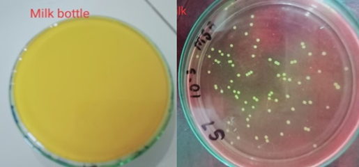
During this study, 50 strains of Staphylococcus aureus were isolated from different food samples obtained from various sources (Table 02). The samples were spread on mannitol salt agar, and the appearance of yellow colonies and change in media coloration indicated S. aureus colonies (Figure 01). Notably, numerous colonies were observed in the milk bottle purchased from Multan, suggesting a potential source of high bacterial contamination. In contrast, food samples from Mailsi City such as shawarma, dahi bhalay, tomato chutney, and green raita tested negative for Staphylococcus aureus. Similarly, samples from Multan, including pizza, fries, potato kebab, cheese halwa, pholky, khoya, and yogurt showed no growth of Staphylococcus aureus. All isolated strains exhibited yellow colonies, confirming their identity as Staphylococcus aureus. Furthermore, all the isolated strains appeared gram positive and cocci in shape on gram staining. Additionally, the isolated strains tested positive for coagulase, catalase, and urease tests, further supporting their identification as Staphylococcus aureus. The Hemolysis assay was performed on the S. aureus strains isolated from different food samples (Figure 02). Strains isolated from Mailsi yogurt exhibited alpha hemolysis characterized by a light greenish color and incomplete breakage of red blood cells. Strains isolated from yogurt samples from other locations revealed a clear zone as red blood cells were completely lysed. There was no clear zone in the case of isolates from chicken, indicating gamma hemolysis. 50 percent of isolates were beta hemolytic, 30 percent were alpha hemolytic and 20 percent were gamma hemolytic.
Biofilm experiments of isolated S. aureus strains were carried out to assess their biofilm-forming capabilities.
Table 2. Isolation and identification of S. aureus from different samples
|
Sample |
Location |
Sample name |
Number of colonies observed |
Number of colonies purified |
|
1 |
Multan |
Cheese halwa |
No growth |
- |
|
2 |
Multan |
Pholky |
No growth |
- |
|
3 |
Multan |
Bakar Khani |
10 |
2 |
|
4 |
Multan |
Chicken |
20 |
6 |
|
5 |
Multan |
Fries |
No growth |
- |
|
6 |
Multan |
Pizza |
No growth |
- |
|
7 |
Multan |
Potato Kebab |
No growth |
- |
|
8 |
Multan |
Milk |
12 |
6 |
|
9 |
Mailsi |
Dahi Bhalay |
No growth |
- |
|
10 |
Mailsi colony chowk |
Shawarma |
No growth |
- |
|
11 |
Mailsi food street |
Shawarma |
No growth |
- |
|
12 |
Mailsi |
Raw Milk |
12 |
7 |
|
13 |
Mailsi |
Green raita |
No growth |
- |
|
14 |
Mailsi |
Tomato chutney |
No growth |
- |
|
15 |
Mailsi |
Yogurt |
6 |
3 |
|
16 |
Mailsi |
Chicken |
10 |
3 |
|
17 |
Multan 6 No. |
Milk |
4 |
2 |
|
18 |
Multan 6 No. |
Chicken |
4 |
5 |
|
19 |
Multan 6 No. |
Milk bottle |
Too numerous to count |
10 |
|
20 |
Multan 6 No. |
Khoya |
No strain |
- |
|
21 |
Multan 6 No. |
Yogurt |
7 |
5 |
Figure 1. Strains of Staphylococcus aureus on mannitol salt agar (yellow)
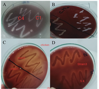
Figure 2. A and B show gamma hemolysis. In C, both strains show alpha hemolysis. In D, strain 2 indicates beta hemolysis and strain 1 exhibited alpha hemolysis
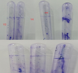
Figure 3. From the yogurt sample, isolate Y3 is a weak biofilm former whereas Y4 is a moderate biofilm former. In Figure B, the strain isolated from the milk bottle shows strong biofilm formation and the second tube shows weak biofilm by an isolate from chicken. In C, the first tube showed weak biofilm of strain isolated from Mailsi milk and the other two showed moderate biofilm of strains isolated from Multan milk and chicken, respectively
Yogurt samples showed a predominance of moderate and weak biofilm production, whereas bacteria isolated from Mailsi milk produced weak biofilm, as shown in Figure 3. On the other hand, isolates of chicken samples in Multan showed moderate biofilm formation, whereas strains from the milk bottle purchased in Multan showed significant biofilm formation. These differences in the biofilm formation patterns highlighted the bacterial potential for pathogenicity. Noteworthy is the prevalence of strong biofilm formers in samples from the Multan milk bottle, suggesting potential sources or conditions contributing to this phenomenon.
3.1 Antibiotic Susceptibility Test
The antibiotic susceptibility test was performed using four antibiotics: vancomycin, ampicillin, erythromycin, and amoxicillin (Table 03). All S. aureus isolated from food samples were found resistant to amoxicillin and ampicillin (Figure 04). Only one strain, designated as Yma and isolated from Mailsi yogurt, showed susceptibility to erythromycin, while other strains exhibited resistance. Variable results were observed while testing the antimicrobial effect of vancomycin, with four strains showing resistance and seven isolated strains showing susceptibility. These findings indicated that the isolated S. aureus strains were multidrug-resistant and highly virulent.
Table 3. Antibiotic susceptibility profiling of S. aureus isolated from food samples
|
Strains |
Vancomycin |
Ampicillin |
Erythromycin |
Amoxicillin |
|
SY1 |
S |
R |
R |
R |
|
SMbot1 |
R |
R |
R |
R |
|
YB |
Intermediate |
R |
R |
R |
|
CM3 |
S |
R |
R |
R |
|
Cm |
Intermediate |
R |
R |
R |
|
Cmu |
Intermediate |
R |
R |
R |
|
Yma |
S |
R |
S |
R |
|
Mma |
Intermediate |
R |
R |
R |
|
Mm6 |
Intermediate |
R |
R |
R |
|
Mm |
Intermediate |
R |
R |
R |
|
MB |
R |
R |
R |
R |
|
Yma2 |
S |
R |
R |
R |
|
YB3 |
S |
R |
R |
R |
|
Mbo1 |
R |
R |
R |
R |
|
Mbo2 |
R |
R |
R |
R |
|
Cma4 |
Intermediate |
R |
R |
R |
|
Chm7 |
S |
R |
R |
R |
|
YB3 |
S |
R |
R |
R |
|
Mm2 |
Intermediate |
R |
R |
R |
*R=resistance and S=sensitive
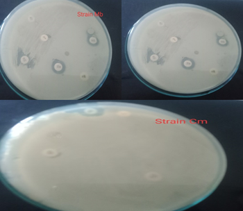
Figures 4. Antibiotic susceptibility testing of S. aureus isolates from food samples
The emergence of antibiotic-resistant S. aureus strains in food samples presents a significant public health concern. In the current study, investigated the antibiotic susceptibility patterns of S. aureus isolated from various food sources and evaluated their potential virulence factors. Findings revealed various valuable insights into the prevalence of antibiotic resistance and the virulence potential of these foodborne pathogens. By testing the different food samples, food hygiene was assessed. Street products showed no growth of S. aureus. Raw food samples like yogurt, milk, and chicken were positive for S. aureus isolates. S. aureus was accompanied by a high degree of virulence and is considered the most virulent pathogen. A total of 50 S. aureus strains were isolated on mannitol salt agar, which is a selective media for S. aureus. The appearance of yellow colonies and the change in the color of the media was an interpretation of positive results. All isolated strains were tested positive for catalase, coagulase, and urease test. Results indicated a high prevalence of multidrug-resistant S. aureus strains among the isolates tested. The rise of multidrug resistance poses challenges to the efficient treatment of S. aureus-related illnesses and emphasizes the urgent need for alternative therapeutic approaches [26].
In this study, 50 strains were evaluated for hemolysin production. 3 types of hemolysins were observed, and results indicated that 50 percent of isolates were beta-hemolytic, 30 percent were alpha-hemolytic, and 20 percent were gamma hemolytic. These varied hemolysis patterns suggested variations in the strain’s potential to rupture red blood cells, which may indicate differences in virulence among the strains [27]. Previous studies have shown that most S. aureus strains isolated from buffalo milk were delta hemolysin producers [28]. While alpha and beta hemolysis was observed by S. aureus strains of human origin. It has been reported that coagulase-positive isolates from animal origin exhibited beta hemolysis [29]. Hemolysins play an important role in increasing the virulence of bacteria. Hemolysin production by S. aureus can make it resistant [30].
Many virulent factors play a crucial role in the pathogenesis of this bacteria allowing it to colonize and persist within the host cell. Several exotoxins are secreted by these bacteria in which a group of polypeptides (gamma hemolysin, alpha hemolysin and phenol soluble modulins) is also included, which can destruct the plasma membrane of the host cell. The hemolytic activity of S. aureus results in the lysis of red blood cells [31]. Alpha hemolysins are also effective against mammalian cells like human red blood cells and can be neurotoxic to humans. A two-component toxin namely gamma toxin is a constituent of 6 different proteins and is actively involved in the breakdown of red blood cells. A low molecular weight toxin namely alpha toxin has the ability to cause lysis of many cell types [32]. A non-pore-forming toxin namely beta toxin is a sphingomyelinase and is involved in hemolysis. Beta toxin is related to the DNase 1 superfamily and there might be a possibility that it causes cleavage of DNA [33].
It has been observed that biofilm formed by microbes is responsible for 80 percent contamination of food. S. aureus is a well-known food-borne pathogen and can form biofilm efficiently, thus is responsible for severe food poisoning and contamination of food processing equipment [34]. Biofilm is a defense system utilized by bacteria to survive in harsh conditions and to make community of cells surrounded by EPS (extracellular polymeric substances). Extracellular DNA (eDNA) is present in biofilm and is considered a crucial component. eDNA enhances the adhesion capacity of bacteria [35]. S. aureus can form biofilm on both living and non-living surfaces. Strains that form biofilm are characterized by higher resistance to antibiotics than those who do not form [36]. Contamination level of food surfaces with S. aureus reveals that the hygienic standard of food industries should be improved and livestock should be handled in a better way. Foods get contaminated due to the biofilm formation and this all happen due to inappropriate use of cleaning agent against bacterial cells forming biofilm [37]. Biofilm associated protein is crucial for the initial adherence of S. aureus and for its biofilm formation [38]. In this study, the Crystal violet staining method was used to test for biofilm formation. The appearance of the ring in the bottom of the tube was an indication of a positive result. Thick ring formation means that the strain exhibits strong biofilm formation. Thin ring formation indicates weak to moderate biofilm formation. Strong biofilm formation was observed by isolates obtained from milk bottles and weak biofilm formation by chicken isolates. Whereas moderate biofilm formation was observed from strains isolated from yogurt. The high tendency for biofilm formation revealed the virulence of strains. The stronger the biofilm formation, the more infectious the strain is [39]. One study revealed that methicillin resistant S. aureus from milk sources exhibited high potency for biofilm formation [40].
Antibiotic resistance can be transmitted through food, and this happens due to the presence of antibiotic residues in food and resistant food pathogens. Most of the commonly used antibiotics are ineffective on Staphylococcus aureus because of an exopolysaccharide barrier which is produced by this bacteria within the micro abscesses thus limiting the activity of drugs [41].Most S. aureus strains were resistant to ampicillin, amoxicillin, and erythromycin in the current study indicating that they were multidrug resistant. All isolated strains exhibited resistance against ampicillin and amoxicillin. Only a few strains from the milk bottle were resistant to vancomycin whereas a few strains were susceptible and some showed intermediate resistance to vancomycin. In a previous study, all test strains were resistant to methicillin but were found sensitive to vancomycin [42].
Overall, the study highlights the consequence of continued surveillance and monitoring of virulence factors and antibiotic resistance in foodborne pathogens to inform public health interventions and mitigate the risk of foodborne illnesses. Future research efforts should focus on elucidating the mechanisms underlying antibiotic resistance and virulence in S. aureus strains isolated from food samples, as well as developing alternative strategies for the control and management of foodborne infections.
In conclusion, this study emphasizes the alarming prevalence of multidrug-resistant S. aureus strains in several food sources, especially from raw foods. So, consumption of raw foods should be avoided as it is a health risk. During baking, hygienic protocols are not properly followed leading to contamination of bakery products. The presence of S. aureus in food can cause food poisoning. There should be proper hygienic control in food industries. Surface sterilization and personnel hygiene of industry workers should be regularly monitored. Food should be cooked properly. Resistance to common antibiotics like ampicillin and amoxicillin was widespread among the isolates, emphasizing the determination for alternative treatment strategies. Variability in susceptibility patterns to other antibiotics was also observed, emphasizing the need for continuous monitoring of antibiotic resistance trends in foodborne pathogens. Furthermore, the presence of diverse biofilm-forming capabilities and hemolytic activity among the isolates emphasizes the complexity of microbial virulence. Strengthening food safety measures and antimicrobial stewardship programs are crucial to mitigate the spread of antibiotic-resistant strains and reduce the risk of foodborne illnesses. Future research should focus on understanding the underlying mechanisms of antibiotic resistance and virulence in S. aureus strains from food samples, facilitating the development of effective interventions to safeguard public health.
REFERENCES