Exploring Skin Microbiota: Insights into Antibiotic Resistance and Environmental Influence
|
1Department of Microbiology and Molecular Genetics, The Women University Multan, Pakistan 2Department of Eastern Medicine, Faculty of Medicine and Allied Health Sciences, The Islamic University of Bahawalpur, Pakistan *Correspondence Humaira Yasmeen Email:humaira.6127@wum.edu.pk |
|
Keywords: Skin Microbia, Antibiotic Resistance, Enviornmental Factors, Public Health, Microbial Diversity |
Hafsa Rehman1 | Humaira Yasmeen1 | Ume Habiba2 | Muhammad Asim2 | Hamza Ishfaq2
|
ABSTRACT Human skin microflora is an important barrier for many harmful invading microorganisms. However, innate and foreign factors including metal exposure and overuse of antibiotics may attune this microflora. Disruption of microflora in turn leads to several skin diseases and other health problems. The current study aimed to isolate antibiotic-resistant microorganisms and explore new methods for treating these microorganisms of skin flora. For this purpose, 100 skin samples were collected from urban and rural agricultural residents of Multan. Bacterial isolates were biochemically characterized. Isolated microflora included S. aureus 24.5%, S.epidermidis (28%), C.xerosis (8.5%), Klebsiella (9%), C. Kutscheri (5%), P.aurigenosa (19.6%), and E.coli (5.5%). |
1. INTRODUCTION
Skin functionally acts as a physical barrier and prevents foreign pathogens invasions. Many intrinsic and extrinsic factors influence microbiota diversity and composition of skin flora. Mainly skin microflora include S. epidermidis, S. aureus and P. acnes [1]. The composition of human skin flora is determined even before birth. Many environmental and host-related factors modulate microbiota structure throughout life. Human skin flora is unique in each individual [2]. The microorganisms of normal microflora are opportunistic, they exist on skin asymptomatically but sometimes they become pathogenic when the host becomes immunocompromised [3]. Culture-based investigations of skin microbiota showed that skin organisms can influence skin properties, microorganism development, and wound recuperation [4].
The rise of antimicrobial obstruction is unavoidable in pretty much every new medication [5]. The use of antibiotics on a mass scale has prompted the rise of the microorganism’s resistance capability [6]. These resistance-causing genes transfer horizontally within same species as well as in different species resulting in extensive multiple drug resistance among different species [7]. In response to high-dose antimicrobial therapy resistance in microbiota shows the development of their evolutionary processes that select genetically and physiologically capable strains that can tolerate the high dose of antibiotics [8]. This increasing antibiotic resistance become a serious alarm of global health. The contracting exhibit of powerful antibacterial medications lifts mortality and expands the recurrence and scale of the epidemic [9].
For all microbial growth aspects, metals act directly or indirectly. Many metals interact with microbial cells and vary their physical and chemical mechanism and their transport system. It directly or indirectly depends on their metabolism. The metals are also toxic to human skin if it is exposed to high levels [10]. The knowledge of skin microbiome is necessary to enhance understanding of human skin disorders and also determine the involvement of skin microflora on skin disorders for approaching new treatments [11]. Moreover, intrinsic and extrinsic host factors also influence the composition of microbiota. Variation in the skin during aging is the main factor mainly correlated with these taxa independently [12].
Present study aims to explore diversity of skin microflora under different stressed environment. It will also explore difference of rural and urban settings on skin flora.
2. MATERIAL AND METHODS
2.1 Sample Collection
Skin specimens were obtained from the urban and rural agricultural areas of Multan. A total of 100 samples were taken with the help of a sterile moist swab from the volar forearms of healthy humans. The ratio of male and female was 1:1. Demographics data was obtained by filling out a questionnaire after their consent.
2.2. Isolation and Characterization of Bacteria
The skin specimens were cultivated on nutrient agar by pour plate method (1ml) and incubated for twenty-four hours at 37 ºC. Colony forming unit (CFU) was calculated for each plate. Bacterial isolates were selected based on their distinct morphology and were processed further for biochemical characterization.
2.3 Biochemical Characterization
After Gram staining, biochemical methods such as Catalase, Oxidase, Triple Sugar Iron (TSI), Citrate, Coagulase, and Indole tests were used to identify bacterial isolates by using 18–24 hours fresh cultures according to Bergey’s Manual of Systematic Bacteriology. In particular, tests for oxidase, glucose fermentation, and Indole were conducted for gram-negative bacteria. In contrast, coagulase, TSI, and catalase tests were run for gram-positive bacteria. Relative abundance (%) within the skin microflora community was used to express the bacterial diversity of the skin microflora.
2.4 Antibiotics Sensitivity Testing
The isolates were tested against 8 different antibiotics using the Kirby-Bauer disc diffusion technique [13]. Antibiotic discs were placed in freshly produced lawns of each isolate on Mueller-Hinton agar (MHA) and incubated at 37°C for 24-48 hours. Diameter of zone of inhibition was measured and classified as resistant, sensitive and/or intermediate.
3. RESULTS
3.1 Sample Characteristics
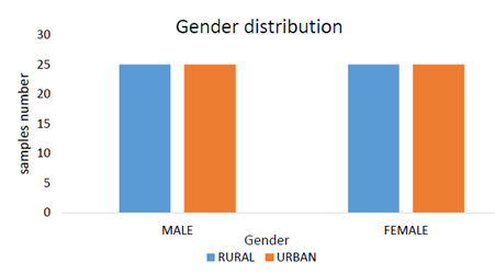
Samples were collected from the volar forearms of 100 healthy volunteers ahead with the directions of a question sheet which comprised questions related to age, gender, blood group, skin type, pet allergy, antibiotic use, weight, and any type of allergy. Volunteers ranged in age from 22 years to 50 years of which 50 were males and 50 were females. These data were collected from industrial (Urban) and agricultural (Rural) areas of Multan. (Fig 1).
3.2 Morphological Characterization and Gram Staining
From 100 samples, 200 isolates were initially biochemically identified. Gram staining revealed that 70 (35%) of 200 strains were gram-negative.
3.3 Biochemical Characterization
Various biochemical tests like the Catalase test, Oxidase test, Triple Sugar Iron (TSI) test, Citrate test, Coagulase test, and Indole test were performed to identify the bacterial isolates using Bergey’s Manual of Systematic Bacteriology. Among Gram Positives, Staphylococcus epidermidis was most abundantly present (29%) while Corynebacterium kutscheri was least abundant (5%). Klebsella spp. was dominantly present in was dominantly present in gram negative isolates (Figure 2).
Figure 1. Gender and area of volunteers
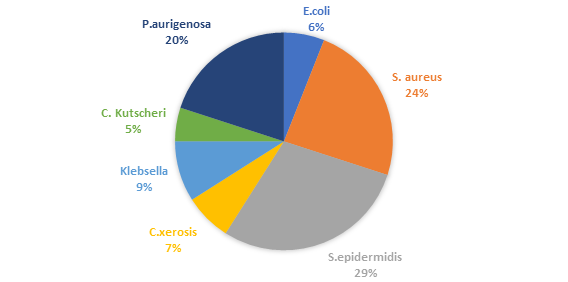
Figure 2. Percentage of isolated strains
S.aureus, C.Kutscheri, P.aeruginosa, and E.coli indicate β-hemolysis (Table 3), which was evident by the absolute crashing of the RBC’s leaving a clear area. The remaining three isolates S. epidermidis, C. xerosis, and Klebsiella showed γ-hemolysis, and no breaking of the RBC’s occurred. In the hemolysis test the ratio of positive and negative was nearly equal (Table 1).
Table 1. Isolated strains' hemolytic activity
|
Strain |
Hemolysis type |
|
S. aureus |
Complete hemolytic |
|
S. epidermidis |
Non-hemolytic |
|
C. xerosis |
Non-hemolytic |
|
Klebsiella |
Non-hemolytic |
|
C.kutscheri |
Complete hemolytic |
|
P. aeruginosa |
Complete hemolytic |
|
E.coli |
Complete hemolytic |
3.4 Antibiotic Susceptibility and Resistance Pattern
The Antimicrobial vulnerability test was done by the disc diffusion method proposed by Kirby Bauer13 conferring to the standards acclaimed by the Muller Hinton agar from the Clinical Laboratory Standards Institute (CLSI). The gram-positive strains of S.aureus, S.epidermidis, C.xerosis, and C. Kutscheri showed susceptibility towards Linezolid, Clindamycin, Oxacillin, Ciprofloxacin, and Gentamycin respectively (Fig 3). Similarly, a trend of resistance was observed by Klebsiella, P.aurigenosa, and E.coli towards Streptomycin, Sulphamethoxazole, and Trimethoprim respectively (Fig 4). S.epidermidis showed maximum zone of inhibition of 19mm, 15mm, 11mm, and 10mm against Linezolid, Clindamycin, Oxacillin, and Ciprofloxacin. On the other hand, all the gram+ve and gram-ve bacteria were resistant to Sulphamethoxazole and Streptomycin (Table 2).
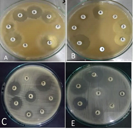
Figure 3. A Staphylococcus aureus, B S.epidermidis, C C.xerosis, E C. Kutscheri. Number 1 is showing the antibiotic disc of Linzeolid, 2 for Clindamycin, 3 for Oxacillin, 4 for Ciprofloxacin, 5 for Gentamycin, 6 for Trimethoprim, 7 for Sulphamethoxazole and 8 for Streptomycin
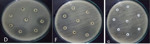
Figure 4. D for Klebsella; F shows P.aurigenosa and G for E.coli. Number 1 is showing the antibiotic disc of Streptomycin, 2 for Sulphamethoxazole, 3 for Trimethoprim, 4 for Ciprofloxacin, 5 for Gentamycin, 6 for Oxacillin, 7 for Clindamycin and 8 for Linezolid
Table 2. Zone of inhibition in mm and resistant pattern of isolated bacterial strains
|
Antibiotics |
Zone of inhibition of isolated bacterial strains |
||||||
|
A |
B |
C |
D |
E |
F |
G |
|
|
Linezolid |
13 |
19 |
13 |
13 |
10 |
7 |
--- |
|
Clindamycin |
12 |
15 |
9 |
10 |
9 |
6 |
--- |
|
Oxacillin |
11 |
11 |
8 |
10 |
7 |
4 |
--- |
|
Ciprofloxacin |
9 |
10 |
8 |
9 |
6 |
3 |
--- |
|
Gentamicin |
8 |
6 |
6 |
8 |
4 |
--- |
--- |
|
Trimethoprim |
6 |
--- |
5 |
5 |
--- |
--- |
--- |
|
Sulphamethoxazole |
--- |
--- |
4 |
--- |
--- |
--- |
--- |
|
Streptomycin |
--- |
--- |
--- |
--- |
--- |
--- |
--- |
A is showing the zone of inhibition and resistance pattern of Staphylococcus aureus, B for S.epidermidis, C for C.xerosis; D for Klebsella , E for C. Kutscheri, F for P.aurigenosa and G for E.coli. --- indicates no zone of inhibition.
4. DISCUSSION
Skin, the largest human body organ is home to numerous microorganisms that have had an impact on one's health. There is a need to quantify the involvement of microbes in healthy human skin at both the community and individual levels. In this study isolated strains ratio of S.epidermis was more than other strains like S. aureus, than other isolated strains. The ratio of Staphylococcus aureus 24.5%, S.epidermidis (28%), C.xerosis (8.5%), Klebsiella (9%), C.Kutscheri (5%), P.aurigenosa (19.6), and E.coli (5.5%). Almost all of the isolates found susceptible to the majority of antibiotics except E. coli. The results of this antibiotic resistance was slightly different from others works According to the Arora and Devi [14] results showed in the company the gram+ve organism’s high resistance was noticed with penicillin (71.4%) which is accordant with other research, where the rate of resistance was 74.61 %. Furthermore, high resistance among gram-ve was seen against penicillin (82.6%), Tetracycline (65.2%). Antibiotic susceptibility showed that isolated strains of gram-negative
showed 100% susceptibility on ciprofloxacin and streptomycin. However, they were showing 100% resistance to clindamycin and oxacillin. The drug trimethoprim sulphamethoxazole they were with efficacy 37.50%, the susceptibility pattern of penicillin and amikacin was with efficacy 25%. The resistance of gram negative was against penicillin was 70% and tetracycline was 74%. Abebaw et al [15] studies showed that the antiseptic susceptibility test for Gram-ve bacteria divulged a higher grade of resistance (>80%) for penicillin (90.4%), and for an intervening grade of resistance to tetracycline (74.2%) and a lower grade of resistance to the last drugs same results are correlated with studies where gram negative bacteria show resistance against antibiotics and proteins extracts of different medicinal plants [16]. Mehdinejad et al [17] studies showed a higher grade of resistance (>80%) against ampicillin and amoxicillin and an intervening grade of resistance to tetracycline and Co-trimoxazol against the antimicrobial sensitivity of Gram-negative bacteria. However, Norfloxacin and ciprofloxacin comparative to other observed drugs are highly sensitive with the precusry research conducted in Jimma and elsewhere in the world.
Ali et al [18] in their studies showed that isolates of Gram-ve and Gram+ve bacteria have ciprofloxacin, vancomycin, ceftriaxone, and norfloxacin were the most potent drug against other isolates that comparable in Ethiopia and elsewhere in the world. This show reflection of the infrequency of prescription of these drugs by physicians and the drugs may be higher cost relative to others but effectiveness of these drugs against the tested organisms So, people do not take these drugs for self-medication.
Worku et al [19] in their study, both Gram +ve and Gram-ve isolates from samples indicate multiple drug resistance. This finding is inconsistent with the previous conducted studies in Ethiopia. The unregulated nonprescription sale of antimicrobials and self-management of an apparent infection in humans without a physician’s instruction. As a results multidrug resistance in Ethiopia lead to the publishing and rapid dissemination of resistant shear. The treatment of bacterial infections are responsible for the increased level of multiple drug resistance with cheaper generic drugs (like amoxicillin) in the market in Jemma town showed that 27.6% of the 152 sick individuals self-medicated. A study on the practice of self-medication. Research revealed that a significant level of resistance to Ampicillin (71.4%) and Penicillin (85.7%) was seen in gram-positive bacteria. There was 100% resistance against Ampicillin, 88.9% to Tetracycline, 77.9% to Chloroamphenicole, 77.8% to Co-trimoxazolee, and 66.7% against Gentamicin against gram-negative bacteria. But resistance for gram +ve was from 0% to 85.7%, and for gram negative from 0% to 100%. Therefore, Ceftriaxone and Ciprofloxacin are more resistant to gram-positive and gram-negative bacterial isolates.
In conclusion, this research has illuminated the complex interplay between skin microbiota, antibiotic resistance, and environmental factors. The findings underscore the importance of holistic approaches in understanding and managing antibiotic resistance, considering both human and environmental dimensions. Moving forward, further research should continue to explore these dynamics to develop targeted strategies for preserving both human health and environmental integrity in the face of rising antibiotic resistance.
REFERENCES