Comparative Analysis of Antibacterial and Antifungal Activity of AgNPs with Conjugated Curcumin AgNPs
Esha Ameen[1]*, Rida Tanveer1, Ayesha Mukhtar[2], Mehreen Fatima2, Muhammad Bilal[3]
|
KEYWORDS AgNPs, Nanoparticles, UV-visible spectrophotometer, FTIR Analysis, Curcumin, Antimicrobial activity |
|
Abstract Silver nanoparticles (AgNPs) are potent antimicrobial agents, extensively used against a wide variety of microorganisms. Several techniques have been developed to chemically synthesize silver nanoparticles but limited their application due to their cytotoxicity and safety concerns for humans and the environment. The current study summarized the preparation of silver nanoparticles from a reaction of silver nitrate with grapefruit extract and to compare the antimicrobial activities of AgNPs and Cur-AgNPs. A natural phenolic compound having mild antimicrobial potential, curcumin was conjugated with initially synthesized silver nanoparticles (Cur-AgNPs) and characterization was performed before and after conjugation by using UV-visible spectrophotometer and Fourier Transform Infrared Spectroscopy (FTIR). The antimicrobial activity of both AgNPs and Cur-AgNPs was assessed against microbial species including gram-positive and gram-negative bacteria. The obtained results led to the conclusion that Cur-AgNPs have more antibacterial and antifungal activity than silver nanoparticles (AgNPs). The antibacterial potential of AgNPs and Cur-AgNPs was evaluated by measuring the diameter of the zone of inhibition in cm. The maximum zone of inhibition measured while using conjugated Cur-AgNPs at a concentration of 0.4mg/uL was 2cm, 1.9cm and 2.2cm against fungus, E.coli and P.aeruginosa respectively. The conjugation of curcumin to silver nanoparticles devised a new biocidal agent and lifted the industrial biomedical application of silver nanoparticles with less toxicity towards the ecosystem.
|
INTRODUCTION
Silver is a noble metal and from early ages, its several compounds have been used for therapeutic and medicinal purposes. Greeks have used it for wound healing and sterilization of instruments [1]. The compounds of silver, silver nitrate and silver sulfadiazine, have shown the benefit in the inhibition of microbial growth. Therefore, they have been used in treating burns, ulcers and wounds to prevent infection [2]. The silver metal and compounds constantly discharge ions on the surface of the metal and work as an antimicrobial [3]. The antimicrobial and antibacterial properties of silver compounds are mainly because of the silver ions. These silver ions react with the thiol groups in the proteins and enzymes of bacteria and other microbes and hinder their respiration and cause cell death [4]. In Pharmacopoeia, in 69 BC, silver nitrate was published as a medicinal compound for the treatment and prevention of wound infections [5]. After several studies on microbial infections in the 1800s, the use of silver as an infection control and antimicrobial increased [6].
Nowadays, nanotechnology is emerging and developing rapidly and is being used in many fields such as biomedical engineering and sciences for the formation of drugs and other chemicals. It deals with the compounds at molecular and atomic levels [7]. Different metals are used as nanoparticles, for example, Cu, Au, Ag and many others. The silver nanoparticles (AgNPs) are well-studied and used for various purposes. They have wide applications in the packaging industry, textile and electronic industries and in pharmaceuticals [8]. The size of silver nanoparticles ranges from 1-100 nm. In the bulk amount, AgNPs are suggested to have more surface area and larger capacity than that of silver metal particles. These nanoparticles have distinct optical, catalytic and electrical properties, therefore, AgNPs are characterized and employed for diagnosis, detection and targeted drug delivery [9]. In addition to these applications, AgNPs gained attention of the scientists and researchers because of their antibacterial activity. AgNPs have shown inhibitory effects on many microorganisms, for example, multidrug-resistant bacteria [10].
Citrus fruits and their products have a wide variety of compounds and pigments in them which are being used in pharmaceuticals and cosmetics. These compounds are of great benefit, as citrus extract has been used to extract vesicles. These vesicles were used for treating cancers and many other diseases [11]. Grapefruit has a very unique chemical composition including polyphenols, tocopherols, essential oils, minerals, phytosterols, carotenoids and many other compounds [12]. It also has many other benefits such as in neuro-protection, improvement of lipid metabolism and regulation of body weight. It has also shown its interaction with drugs and acts as immunosuppressants, blocking agents and antihistamines [13].
Many studies have suggested that the peels of citrus fruits have been used for the formation of nanoparticles of silver, iron oxide, zirconium, zinc oxide and titanium oxide [14]. Industrially, the plasma discharge method has been employed to prepare these nanoparticles. Many methods were used to modify the nanoparticles including photolysis reaction, UV radiation and other redox reactions. These methods increase the concentration and efficiency of the yield of nanoparticles [15].
Curcumin is the most prominent component of the polyphenols. It is extracted from a rhizome plant called turmeric (scientifically known as Curcuma longa) [16]. Curcumin works as an anti-inflammatory agent. The immune response increases the production of inflammatory molecules including reactive oxygen species, cytokines, nuclear factor kB and cyclooxygenases. Curcumin weakens the immune response and stabilizes the levels of these molecules [17][18]. Besides anti-inflammation, it also has numerous other benefits such as antibacterial, antifungal and antiviral activities, metabolic and immune regulation, tissue protection, anti-depressant, anticancer and antioxidant properties [19][20].
A study has shown that high levels of curcumin were found in the gastrointestinal tract after oral intake.[21]. It was hypothesized that high levels of curcumin regulated the growth of gut microbiota. It has high pharmacological activity but low bioavailability [22][23]. To increase the bioavailability of curcumin, it is conjugated with nanoparticles. These conjugated nanoparticles are used as therapeutic agents to deliver curcumin. Hence, it has a great benefit in targeted drug therapies [24]. Curcumin is encapsulated in nanoparticles which enhance the stability of curcumin and prevent it from degradation by enzymes. It also helps in its circulation in the body [25][26].
Silver nanoparticles along with curcumin were observed to inhibit the bacterial growth and formation of biofilms. It has shown high antibacterial activity against the gram-negative bacteria, E.coli [27]. The silver nanoparticles are being characterized with the help of many techniques including spectroscopy and antimicrobial testing [28]. Majorly, UV-visible spectroscopy is being employed to identify the production and size of nanoparticles [29]. Other citrus fruits have been used before but grapefruit was selected because of its health benefits. In this study, nanoparticles are formed with the help of grapefruit extract. AgNPs and curcumin conjugate with AgNPs (Cur-AgNPs) were characterized using UV-visible spectroscopy and Fourier transform infrared spectroscopy (FTIR). Further antibacterial and antifungal activities of AgNPs and cur-AgNPs were observed and analyzed the efficiency of AgNPs and cur-AgNPs.
MATERIALS AND METHODS
Merck’s silver nitrate base was used and grapefruit extracts were reduced and stabilized. The entire solution was prepared in double distilled water. The grapefruits of premium quality were selected from the local market. These were washed with distilled water, squeezed mechanically, and kept in the glass jars for storage. Antimicrobial and antibacterial activities were observed and studied by using microbial and bacterial cultures. Curcumin-silver nanoparticles were made using the chemical called curcumin.
Formation of Silver Nanoparticles (AgNPs) from Grapefruit Extract
The grape fruit extract has a distinct sour taste which is because of the presence of acid. Firstly, the grapefruit extract was extracted by employing simple and efficient methods.
Conjugation of Curcumin with AgNPs (Cur-AgNPs)
Curcumin powder was prepared for making the conjugated cur-AgNPs. For the formation of cur-AgNPs, 7mg of curcumin was dissolved in 20 ml of PEG (polyethylene glycol) to increase the solubility and stability of curcumin in solution. This solution was stirred with the help of an ultrasonicator for 15 minutes at room temperature. Afterward, 20 ml of PEG was taken in the flask and placed on the hot plate. Now, 1g of cur-AgNPs was added to it. A magnetic stirrer was placed in the flask and again kept on the hot plate. 1g of cur-AgNPs, 200 µl of silver nitrate solution, and 10M PVP (polyvinylpyrrolidone) to the flask containing PEG to encapsulate the conjugated cur-AgNPs. This solution was continuously stirred on the hot plate for 10 minutes.
Characterization of AgNPs and Cur-AgNPs
For the characterization of the silver nanoparticles (AgNPs), several different methods were suggested and performed including spectroscopy, Fourier transforms infrared spectroscopy (FTIR), X-ray diffraction, Scanning Electron Microscopy (SEM), Dynamic Light Scattering (DLS), etc. These techniques were used for the verification of the AgNPs production. In addition, these techniques described the actual size of these nanoparticles. In the given study, UV-visible spectroscopy and FTIR were used for the characterization of silver nanoparticles.
UV Visible Spectroscopy
Spectroscopy is described as absorption spectroscopy in which the spectral region lies within the ultraviolet and visible regions. It is a widely suggested method for verifying the structural integrity of AgNPs.
FTIR Analysis
Fourier transforms infrared spectroscopy (FTIR) was used for further analysis. It gives detailed information about the functional organizations of the nanoparticles by evaluating the vibrational patterns of the chemical interaction and bonds. It helps in the in-situ examinations of interfaces and determines the adhesion of the functional group atoms to the surface of the nanoparticles.
Determination of Antibacterial Activity
1ml of AgNPs suspension was added to each flask and also distilled water was added in proper supervision. Further, after regular intervals, the aliquots of every culture were taken for the measurement of O.D. The growth curves of different strains were made by plotting values of O.D of every culture against time. It was observed that the O.D of both cultures gradually increased and showed fast growth in the absence of AgNPs. On the contrary, bacterial growth decreased in the presence of AgNPs.
Determination of Antifungal Activity
RESULTS
Preparation of Silver Nanoparticles (AgNPs) from Grapefruit Extract
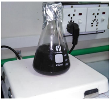
During this technique, the black color of the solution formed because of an excitation state when (AgNPs) vibrated at the surface, Plasmon displayed the formation of silver nanoparticles (AgNPs) from grapefruit extract.
Figure 1. Formation of silver nanoparticles (AgNPs) from grapefruit extract
Microscopic View of Silver Nanoparticles (AgNPs)
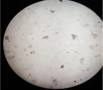
Figure 2. Microscopic view of silver nanoparticles (AgNPs)
Fluorescent Microscopy of Silver Nanoparticles (AgNPs)
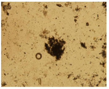
Figure 3. Silver nanoparticles (AgNPs)
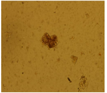
Figure 4. Conjugated curcumin-AgNPs
UV-visible Spectroscopy Analysis on AgNPs
Table 1. UV-visible spectroscopy on AgNPs
|
Treatments |
Wavelength |
Absorbance |
|
T0 |
300 |
1.1 |
|
T1 |
320 |
1.5 |
|
T2 |
340 |
2.0 |
|
T3 |
360 |
2.25 |
|
T4 |
380 |
2.45 |
|
T5 |
400 |
2.75 |
|
T6 |
420 |
3 |
|
T8 |
460 |
2.2 |
|
T9 |
480 |
1.5 |
|
T10 |
500 |
0.9 |
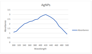
Figure 5. Ultraviolet visible spectroscopy analysis on AgNPs
The development and durability of silver nanoparticles (AgNPs) were examined along with absorption spectra of AgNPs solution using ultraviolet visible spectroscopy when AgNPs were present in colloidal solution. Ultraviolet visible spectroscopy indicated a maximum absorbance of 3.0 at a wavelength of 420 nm. In the figure, curves were assessed for different absorbance ranges at different wavelengths.
UV-visible Spectroscopy Analysis on Conjugated Cur-AgNPs
The conjugated Curcumin -AgNPs were prepared by using PEG as a stabilizing agent through sono chemical reaction. During this conjugation process, curcumin solution color changed from a light yellow color into a pale flash yellow color with a successive transition to deep yellowish pointing out the reduction of silver ions by curcumin. At a wavelength of 300-500 nm, UV-visible spectroscopy analysis revealed that Curcumin nanoparticles enhanced the absorption and stability of silver nanoparticles (AgNPs) as conjugated Cur-AgNPs. These conjugated particles gave maximum absorbance at a wavelength of 430 nm as shown in the figure.
Table 2. Ultraviolet-visible spectroscopy on conjugated Curcumin-AgNPs
|
Treatments |
Wavelength |
Absorbance |
|
T0 |
350 |
0.45 |
|
T1 |
360 |
0.55 |
|
T2 |
370 |
0.65 |
|
T3 |
380 |
0.73 |
|
T4 |
390 |
0.87 |
|
T5 |
400 |
1 |
|
T6 |
410 |
1.3 |
|
T7 |
420 |
1.5 |
|
T8 |
430 |
1.57 |
|
T9 |
440 |
1.25 |
|
T10 |
450 |
0.3 |
|
T11 |
460 |
0.25 |
|
T12 |
470 |
0.2 |
|
T13 |
480 |
0.2 |
|
T14 |
490 |
0.15 |
|
T15 |
500 |
0.1 |
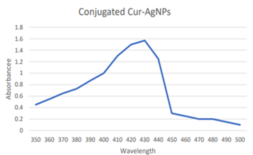
Figure 6. UV-visible spectroscopy analysis on conjugated Curcumin -AgNPs
Fourier Transform Infrared Spectroscopy (FTIR) Analysis of AgNPs

Figure 7. FTIR of Silver nanoparticles (AgNPs)
FTIR analysis revealed maximum absorption at a wavelength of 1636 cm-1 when silver ions were removed to observe feasible biomolecules. This absorption band may represent a C=C double bond or a C-O bond.
Fourier Transform Infrared Spectroscopy (FTIR) Analysis of Conjugated Cur-AgNPs
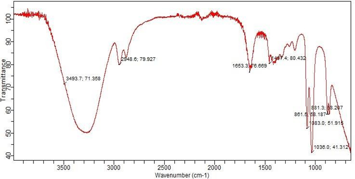
Figure 8. FTIR of conjugated Curcumin-Silver nanoparticles (Cur-AgNPs)
The conjugation of curcumin with AgNPs with characterization of Cur-AgNPs was examined through FTIR analysis. The peaks through FTIR analysis revealed that Cur-AgNPs have some distinct functional groups and intermolecular bonding. The spectrum of analysis indicated the presence of aromatic carbon-hydrogen bonding, Nitrogen-Hydrogen, and Oxygen-Hydrogen carboxylic acid, Carbon-Oxygen-Carbon stretch, Carbon-Nitrogen stretching of an aromatic amine group, and Carbon-Nitrogen stretching ester, and others in the residual solution and NO3- group, Carbon=Carbon bonding, Carbon-Hydrogen stretch at a wavelength of 1036, 1083, 1451, 1653, 2948, and 3493 cm-1 respectively. This collection of various functional groups originated from heterocyclic elements of curcumin that were generally dissolved in water.
AgNPs and Conjugated Curcumin-AgNPs act as Growth Inhibitors
Silver nanoparticles (AgNPs) extracted from grapefruit extract when conjugated with curcumin inhibit the growth of a number of microbes. However, silver nanoparticles (AgNPs) also have the potential for growth inhibition without conjugation. Our research mainly focuses on the growth inhibition of fungi, Escherichia coli and Pseudomonas aeruginosa by assessing antimicrobial activity of conjugated Cur-AgNPs and AgNPs.
In contrast to silver nanoparticles, conjugated Cur-AgNPs have more antifungal and antibacterial activity as shown below by measuring the diameter of the zone of inhibition after the application of AgNPs and conjugated Cur-AgNPs individually on microbial growth. Curcumin itself is unstable and readily degradable but when combined with silver nanoparticles, it becomes photostable thus contributing to its antioxidant and antimicrobial properties.
Comparative Analysis of AgNPs and Conjugated Curcumin-AgNPs
The antifungal property of AgNPs and conjugated Cur-AgNPs was calculated on agar-plate cultures by measuring the diameter of the zone of inhibition (cm) around each well (Table 3). The results demonstrated that AgNPs have less antifungal activity than conjugated Cur-AgNPs against fungi. Using AgNPs as a growth inhibitor at concentrations 0.1mg/uL, 0.2mg/uL, 0.3mg/uL, 0.4mg/uL have 0.5cm, 0.4cm, 0.8cm and 0.9cm diameters of zone of inhibition respectively. However, using conjugated Cur-AgNPs as a growth suppressor at concentrations 0.1mg/uL, 0.2mg/uL, 0.3mg/uL, 0.4mg/uL have more diameter of zone of inhibition 1cm, 0.8cm, 1.5cm and 2cm respectively.
Table 3. Antimicrobial activity of AgNPs and conjugated Cur-AgNPs by measuring zone of inhibition in fungus
|
Concentration |
Conjugated Cur-AgNPs |
AgNPs |
|
0.1mg/µL |
1cm |
0.5cm |
|
0.2mg/µL |
0.8cm |
0.4cm |
|
0.3mg/µL |
1.5cm |
0.8cm |
|
0.4mg/µL |
2cm |
0.9cm |
The effective inhibition of fungi through conjugated Cur-AgNPs is due to the presence of Curcumin as it has the ability to disrupt membrane integrity while silver nanoparticles stop cell division leading to cell death. When silver nanoparticles (AgNPs) are conjugated to Curcumin, silver ion reduction occurs which elevates its antifungal property.
Comparatively Analysis of Antibacterial Properties of AgNPs and Conjugated Cur-AgNPs
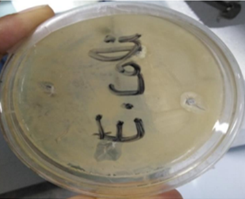
Figure 9. Antibacterial activity of conjugated Cur-AgNPs on E.coli
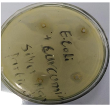
Figure 10. Antimicrobial activity of AgNPs on E.coli
Table 4. Antibacterial activity of AgNPs and conjugated Cur-AgNPs by measuring zone of inhibition in E.coli
|
Concentration |
Conjugated Cur-AgNPs |
AgNPs |
|
0.1mg/µL |
1.2cm |
0.5cm |
|
0.2mg/µL |
1.5cm |
0.8cm |
|
0.3mg/µL |
1.8cm |
0.9cm |
|
0.4mg/µL |
1.9cm |
1.2cm |
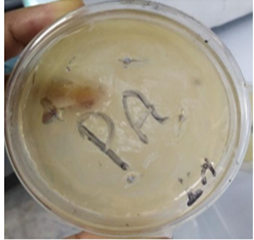
Figure 11. Antibacterial activity of AgNPs on Pseudomonas aeruginosa
Table represents the antibacterial effects of AgNPs and conjugated Cur-AgNPs on E.coli. When AgNPs and conjugated Cur-AgNPs were inoculated at a concentration of 0.1mg/uL-0.4mg/uL each on two separate agar plates, both showed zones of inhibition (cm) around each well as shown in the figure. The comparative analysis depicted that when AgNPs are conjugated with curcumin, enhanced its antibacterial activity, thus increasing the diameter of the zone of inhibition by decreasing the number of E.coli cells (Table 4).
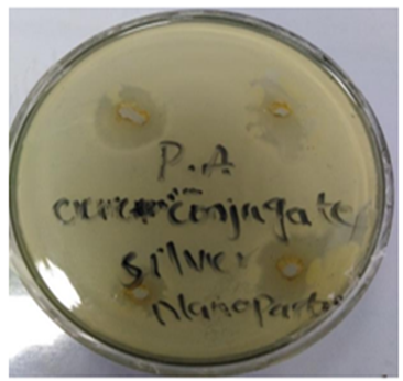
Figure 12. Antibacterial activity of conjugated Cur-AgNPs on Pseudomonas aeruginosa
Table 5. Antibacterial activity of AgNPs and conjugated Cur-AgNPs by measuring zone of inhibition in Pseudomonas aeruginosa
|
Concentration |
Conjugated Cur-AgNPs |
AgNPs |
|
0.1mg/µL |
1.3cm |
0.6cm |
|
0.2mg/µL |
1.6cm |
0.8cm |
|
0.3mg/µL |
1.9cm |
1cm |
|
0.4mg/µL |
2.2cm |
1.2cm |
By using the cell diffusion method, the antibacterial activity of AgNPs and conjugated Cur-AgNPs was also calculated by measuring the zone of inhibition (cm) in Pseudomonas aeruginosa. The table 5 specified that conjugated Cur-AgNPs were found to have higher antimicrobial activity against P. aeruginosa as compared to the antimicrobial activity of AgNPs.
From these results, it is implicated that silver nanoparticles when combined with curcumin, have a synergistic effect eradicating a broader spectrum of microorganisms like bacteria and fungi [30]. Curcumin has anti-inflammatory and anticancer agent properties that inhibit tumorigenesis while silver nanoparticles also have cytotoxic effects. These nanoparticle- anticancer drugs have dual action against cancerous cells and can be used as a therapeutic agent to treat human pathologies in the future [31]. Furthermore, these particles can also be used as biosensors for detecting specific biomolecules [32].
DISCUSSION
Silver nanoparticles are powerful biocidal agents that bind preferentially to sulfur-containing proteins and phosphorus-containing DNA inside the cell or the cell membrane of the microorganism [33]. The formation of pores occurs when silver nanoparticles bind to the phospholipid layer of the bacterial cell membrane, resulting in destabilization and increased membrane permeability [34]. Moreover, these particles can bind to protein in the cell membrane or active center of the enzyme leading to the generation of reactive oxygen species (ROS) inside the bacteria that ultimately result in cell death [35]. A natural phenolic compound isolated from turmeric spice; curcumin has an antibacterial effect against a wide variety of bacteria. The antimicrobial action of curcumin involves damage to cell wall, cell membrane, bacterial quorum sensing, and bacterial growth and causes increased lipid peroxidation and DNA fragmentation in E.coli [36][37]. Curcumin could lessen the expression of 31 quorum-sensing genes and inhibit multiple virulence factors and biofilm formation detected through genome microarray analysis of Pseudomonas aeruginosa. It also inhibits the FtsZ accumulation of Bacillus subtilis and Escherichia coli thus inhibiting cell proliferation [27].
Our research indicated an agreement with the previous research in which Jaiswal and Mishra suggested that curcumin alone is able to reduce and stabilize silver nanoparticles when conjugated Cur-AgNPs are synthesized in the 25–35 nm size range. These conjugated particles serve as an active microbial agent with long period activity and a Minimum Inhibitory Concentration (MIC) of 5 µg/mL [38]. In a study, it was suggested that curcumin conjugated silver nanoparticles (Cur-AgNPs) are synergistic antibacterial particles in which curcumin acts as a reducing and capping agent. These particles at low concentration have a much stronger ability to stop the growth of bacteria. With the help of curcumin, the binding of Ag to bacterial membrane increase and Ag+ release which cause a temporary high Ag+ concentration near the surface of the bacterium, leading to the generation of ROS which ultimately damages membrane and lipases followed by cell death [28]. In another study, Gupta et al., 2020 synthesized silver nanomaterial utilizing curcumin-cyclodextrins to apply them in the wound healing process. These composites exhibit antibacterial action against three wound infection-causing bacteria Staphylococcus aureus, Pseudomonas aeruginosa, and Candida auris [39].
The study emphasizes a comparative analysis of the antimicrobial activity of silver nanoparticles (AgNPs) from grapefruit extract and conjugated curcumin conjugated silver nanoparticles (Cur-AgNPs) synthesized eco-friendly. Characterization through UV-visible spectrophotometer revealed that conjugated Cur-AgNPs gave maximum absorption at 430mm and AgNPs gave maximum absorption at 420 nm while Fourier Transform Infrared Spectroscopy (FTIR) indicated the existence of distinct functional groups and intermolecular bonding in conjugated Cur-AgNPs. The antimicrobial potential of both AgNPs and Cur-AgNPs against three species: fungus, Escherichia coli and Pseudomonas aeruginosa was determined by measuring zone of inhibition on agar culture plates. It is suggested that Cur-AgNPs have a greater antimicrobial effect by giving a maximum zone of inhibition against microbial species than AgNPs because curcumin when conjugated when silver nanoparticles, enhances its biocidal effect and becomes more photostable.
In the future, with the assistance of these nanomaterials we could overcome many pathogenic bacteria involved in different human pathologies like dental problems caused by Streptococcus mutans and nosocomial pneumonia caused by Pseudomonas aeruginosa and used to heal infectious wounds.
LIMITATIONS
Silver nanoparticles are cytotoxic and can accumulate in certain tissues depending upon the size and concentration of silver nanoparticles. Researchers are currently working on it to ensure its bioavailability. Silver nanoparticles need to be stabilized properly to ensure they cannot be released into the environment. There is a need to optimize the formulation process of conjugated curcumin-silver nanoparticles according to the application when using it as an anticancer or antimicrobial agent. Curcumin is degradable and silver nanoparticles whenever exposed to light generate ROS (reactive oxygen species) and produce toxicity.
CONCLUSION
Silver nanoparticles are commonly used as potent antimicrobial agents for the treatment of a broad range of microorganisms. They have prepared through grapefruit extract by utilizing green synthesis strategies rather than chemical methods to reduce environmental pollution. The curcumin contains excellent biologically active compounds which can be used for therapeutic purposes. The silver nanoparticles are conjugated with Curcumin (Cur-AgNPs) and stabilized by it, while curcumin ensures the bioavailability of silver nanoparticles thus inhibiting the growth of many microorganisms due to the enhanced antimicrobial action of Curcumin with silver. The conjugated curcumin-silver nanoparticles performed their synergistic antimicrobial function against both gram-positive and gram-negative strains and hence can be used for the treatment of different microbial infections and cancer.
REFERENCES
*Correspondence: eshaameen974@gmail.com
[1]School of Biological Sciences, University of the Punjab, Lahore, Pakistan
[2]Department of Life Sciences, University of Management and Technology, Lahore, Pakistan
[3]Institute of Scientific and Industrial Research (SANKEN), Osaka University, Japan