
Molecular Fingerprinting and Therapeutic Potential of Two Jasmine Species - Jasminum sambac and Jasminum grandiflorum
Haiqa Saleem, Fatma Hussain, Rafia Rehman, Iqra Saleem, Nadia Nazir
1Department of Biochemistry, Faculty of Sciences, University of Agriculture, Faisalabad, Pakistan
2Department of Biological Sciences, National University of Medical Sciences, Rawalpindi, Pakistan
*Correspondence
Fatma Hussain
Email: fatmauaf@yahoo.com
|
ABSTRACT Jasminum sambac and Jasminum grandiflorum, two jasmine species, have diverse applications in the pharmaceutical and cosmetic industries. This study was designed to assess the antioxidant, antidiabetic, bactericidal, and hemolytic properties of aqueous leaf extracts of Jasminum sambac and Jasminum grandiflorum. The antioxidant activity, total phenolic content (TPC), and total flavonoid content (TFC) were evaluated using DPPH scavenging assay and other recognized protocols. The antidiabetic potential was evaluated through α--amylase inhibition assay (hypoglycemic effect). Hemolytic and bactericidal activities were investigated using hemolysis and well diffusion assays. Furthermore, molecular fingerprinting was performed using Fourier Transform Infrared (FT-IR) spectroscopy. TPC was found to be 30.64 ± 2.96 mg GAE/100 g and 28.28 ± 3.73 mg GAE/100 g for J. sambac and J. grandiflorum, respectively, with no significant difference between the two species (p>0.05). However, J. grandiflorum had a significantly higher TFC (163.93 ± 6.46 mg CE/100 g) compared to J. sambac (144.30 ± 9.55 mg CE/100 g) (p<0.05). Similarly, J. grandiflorum exhibited higher DPPH free radical scavenging activity (58.31 ± 0.24%) compared to J. sambac (47.22 ± 4.65%) (p<0.05). It showed negligible bactericidal activity against S. aureus and mild activity against E. coli. The J. sambac and J. grandiflorum extracts exhibited α-amylase inhibition of 39.26% and 34.66%, respectively. J. sambac and J. grandiflorum exhibited 8.24% and 1.55% hemolysis, respectively. Molecular fingerprinting (FTIR) revealed the presence of alcohols, carboxylic acids, phenols, alkanes, alkenes, esters, ethers, amides, amines and, aromatic compounds. These results underscore the therapeutic attributes of J. sambac and J. grandiflorum leaves, providing valuable insights into their phytochemical makeup and potential health benefits.
|
A significant genus of flowering plants is jasmine, which belongs to the familyOleaceae. Due to its frequent usage in fragrances, tea, sweets, desserts, aromatherapy, skin and hair care products, the Jasmine sambac or Arabic jasmine possesses the utmost economic worth. Its flowers are used in festivals and ceremonies. J. sambac flowers are employed in traditional Asian medications to treat a variety of ailments [1]. However, the ancient herbal remedies made considerable use of Jasminum grandiflorum or Spanish jasmine which is associated with numerous therapeutic benefits, including thermogenic, aphrodisiac, deobstructive, antibacterial, odontalgic, emollient, anthelmintic, and tonic properties. J. grandiflorum is used in traditional medicine for overall well-being and to treat stomatitis, leprosy, dermatitis, ulcers, and calluses. It is also used to reduce pathologies like otalgia [2]. While Jasminum sambac contains numerous primary and secondary metabolites like lipids, essential oils, phenols, alkaloids, terpenoids and alcohols etc.
These metabolites contribute to a wide range of therapeutic effects such as antimicrobial, insecticidal, antipyretic, analgesic, anti-inflammatory, antioxidant, anti-diabetic, anticancer, skin-protective, heart-protective, peroxidation-preventing, and antiobesity properties, as well as gastro-protective effects [3]. Jasminum grandiflorum is particularly noted for its medicinal uses. Most compounds found in its leaves have potential as antioxidants, antimicrobials, anti-inflammatory, antivirals, anti-ulcer, and pain killing agents/ analgesics [4].
Although both jasmine species are well-studied, some aspects have not been unlocked scientifically, such as detailed chemical structure, functional groups, types of bonding, clinical and therapeutic efficacy applications (digestive enzyme inhibition, antiradical, bactericidal), potential side effects and safety assessments (hemolysis), that are frequent challenges in medical conditions like diabetes mellitus, infectious diseases, hepatic disorders and anaemia. Hence, the current study was designed to assess the chemical composition and therapeutic attributes of two jasmine species: Jasminum sambac and Jasminum grandiflorum, to fill this knowledge gap and to gain insight into the potential application in the medicinal landscape.
The powdered leaf samples were mixed with distilled water (1:5 ratio) and heated in a water bath at 45-55 oC for three days. This process was completed with shorter extraction time per day under sterile conditions to minimize contaminations. After filtration, the semi-solid extracts were stored for further analysis [5].
For the evaluation of total phenolic content (TPC), 100 µL of Na2CO3 solution, 0.125 mL of samples, and 0.025 mL of Folin Ciocalteu reagent (diluted to 10%) were mixed, incubated and the optical density was measured at 765 nm. Gallic acid was used as the reference compound and TPC was expressed as mg GAE/100 g [6]. For total flavonoid content (TFC) estimation, 40 µL of sample, 10.5 µL of sodium nitrite and 160 µL of distilled water were mixed. Later, 20 µL aluminum chloride (10%) was added and the mixture was incubated for five minutes. Light absorbance was measured at 510nm. The results were expressed as mg CE/100 g [7]. Antioxidant capacity was evaluated based on its DPPH radical scavenging activity. Briefly, a mixture of 0.25 mL of DPPH solution and 0.025 mL of sample extract was incubated for 35 minutes [7]. Absorbance (A) was recorded at 517 nm. Ascorbic acid was served as the control (0).
2.3 Bactericidal Activity
The antibacterial properties were evaluated using the diffusion method. For sample preparation, 0.5 g of semi-solid aqueous extract was mixed with 5 mL of distilled water. For the agar solution, 2.66 g of agar was dissolved in 70 mL of water and autoclaved along with the Petri dishes. Strains were procured from the Institute of Microbiology, UAF. The inoculum consisted of Staphylococcus aureus (soy broth) and Escherichia coli (LB medium) strains, which were added to the agar solution at 40°C. The agar solution containing bacteria was poured into Petri dishes, and wells were created using a 1 mL micropipette tip. McFarland standard was used for inoculum standardization. Each plate received 80 µL of the sample in three separate wells, along with ciprofloxacin as a positive control. After incubation at 35-37 °C for 16-18 hours, the diameter (mm) of the inhibitory zones (DIZ) was measured in millimeters [8].
2.4 Anti-diabetic Activity - Alpha Amylase Inhibition Assay
Briefly, 30 µL of the sample and 10 µL of alpha amylase solution were mixed. After 10 minutes, starch (1%, 40 µL) was added and the mixture was then incubated for 30 minutes. The HCl (1M, 20 µL) and 75 µL of iodine solution were added. In the negative control, distilled water was used instead of the sample and acarbose was the positive control [8]. The absorbance was measured at 580 nm. To calculate the percentage enzyme inhibition the following formula was used:

2.5 Hemolysis (Anucleated Cells Lysis) Potential
In the hemolytic assay, 3 mL of human blood was taken, centrifuged, and washed with cold PBS (phosphate buffer saline). Then cell suspension, 180 µL of red blood cells (RBCs) and 20 µL of the sample were mixed. After 5 minutes of centrifugation, the liquid phase (100 µL) was diluted with 900 µL of cold PBS. The light absorbance measured at 576 nm was used for calculations. Positive control was Triton X-100 and the negative control was PBS [9]. The percentage of hemolysis was calculated using the following formula:

2.6 Molecular Fingerprinting - Fourier Transform Infrared Spectroscopy (FTIR)
Using a Bruker Tensor 27 FTIR spectrometer, the molecular composition was calculated. The KBr (potassium bromide) and plant extract were mixed and pelletized under high pressure. The FTIR spectra was recorded in a range of 400–4000 cm−1 [10].
2.7 Statistical Analysis
The results of triplicate assessments for each parameter were presented as mean ± SD. To assess the significance of the results between the treatment groups, analysis of variance (ANOVA) was performed using Minitab statistical software version 17.
3.1 Chemical Profiling
Table 1 summarizes the comparative analysis of both species, J. sambac and J. grandiflorum for total phenols, total flavonoids, antioxidant, antidiabetic (alpha amylase inhibition), cytotoxic (hemolysis) and antimicrobial activities. The leaves of J. sambac and J. grandiflorum exhibited almost equal TPC values of 30.64 ± 2.96 mg GAE/100g and 28.28 ± 3.73 mg GAE/100 g, respectively (p > 0.05). In contrast, TFC in J. grandiflorum sample was significantly higher (163.93 ± 6.46 mg CE/100 g) as compared to those of J. sambac (144.30 ± 9.55 mg CE/100 g) (p < 0.05). J. grandiflorum exhibited higher DPPH free radical scavenging activity (58.31 ± 0.24%) compared to J. sambac (47.22 ± 4.65%) (p < 0.05). The IC50 value for J. grandiflorum was 43.10 µg/mL, but for J. sambac it was 53.19 µg/mL.
3.2 Bactericidal Activity
Table 1. Total Phenols, Total Flavonoids, Antioxidant, Antidiabetic (alpha amylase inhibition), Cytotoxic (hemolysis) and Antimicrobial Activities
|
Sample |
Yield (%) |
Total Phenols (mg GAE/100 g) |
Total Flavonoids (mg CE/100 g) |
Antioxidant Activity (%) |
Alpha Amylase inhibition (%) |
Hemolysis (%) |
Antimicrobial Activity (mm) |
|
|
E. coli |
S. aureus |
|||||||
|
J. sambac |
62.26 |
30.64 ± 2.96 |
144.30 ± 9.55 |
47.22 ± 4.65 |
39.26 ± 3.89 |
8.24 ± 0.22 |
9 |
2 |
|
J.grandiflorum |
68.53 |
28.28 ± 3.73 |
163.93 ± 6.46 |
58.31 ± 0.24 |
34.66 ± 0.24 |
1.55 ± 0.05 |
9 |
2 |
|
Control |
- |
- |
- |
89.64 ± 3.71 |
81.47 ± 2.32 |
94.87 ± 1.6 |
27 |
27 |
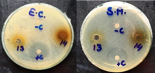
Figure 1. Antimicrobial activity of J. sambac (13) and J. grandiflorum (14) against E.C.: E. coli and S.A.: S. aureus
3.3 Anti-diabetic Activity
The aqueous extracts of J. sambac and J. grandiflorum exhibited anti-diabetic activity by inhibiting the alpha-amylase enzyme, with J. sambac showing 39.26% inhibition and J. grandiflorum showing 34.66% inhibition. Although J. sambac had a slightly higher inhibitory effect, the difference between the two extracts was not statistically significant (p > 0.05). For comparison, the positive control demonstrated 81.47% enzyme inhibition (Table 1).
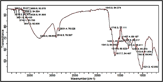
Figure 2. FTIR spectra of Jasminum sambac
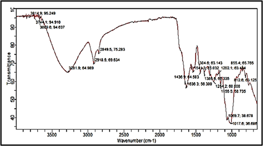
Figure 3. FTIR spectra of J. grandiflorum
Table 2. FTIR Spectral Analysis for the Identification of Major Functional Groups
|
Jasminum sambac |
Jasminum grandiflorum |
||||
|
Peak |
Group |
Compound |
Peak |
Group |
Compound |
|
3854.1, 3839.2, 3801.9, 3744.1 |
O-H |
Alcohol |
3814.9, 3744.1 3669.6, 3281.9 |
O-H |
Alcohol |
|
3647.2 |
O-H |
Alcohols, phenols |
2918.5 |
C-H O-H |
Alkanes (stretch) Carboxylic acids |
|
3289.4 |
O-H |
Alcohols, phenols, Carboxylic acids |
2849.5 |
C-H |
Aldehyde |
|
2851.4 |
C-H |
Alkyne |
1636.3 |
C=C, C=O |
Alkene, Amide |
|
2918.5 |
O-H |
Carboxylic acids |
1541.3 |
C=C, N=O |
Aromatic Nitro (R-NO2) |
|
1845.0 |
C-H |
Aromatic |
1436.9 |
C-H N=O |
Alkanes-CH3 Nitro (R-NO2) |
|
1718.3 |
C=O |
Ketone Carboxylic acid |
1386.6 |
C-H N=O |
Alkanes-CH3 (bend) Nitro (R-NO2) |
|
1617.7 |
N-H |
Amines, amides |
1304.6 |
S=O |
Sulfones, sulfonyl chloride, sulfate, sulfonamides |
|
1541.3, 1457.4 1436.9 |
C=C N=O |
Aromatic Nitro (R-NO2) |
1254.2, 1202.1 1155.5, 1069.7 1017.6 |
C-O C-N S=O C-X |
Ethers, esters, Amines carboxylic acids, anhydrides Sulfones, chloride, sulfate, sulfonamides, Fluoride |
|
1420.1, 1375.4 |
N=O C-H |
Nitro (R-NO2) Alkanes-CH3 |
855.4, 812.6 |
C-H C-H |
Alkenes, Aromatics |
|
1315.8, 1248.8 1153.6 |
C-N S=O C-X |
Amines, Fluoride Sulfones, chloride, sulfates, sulfonamides |
- |
- |
- |
|
780.9 |
C-X C-H C-H |
Chloride Alkenes, Aromatics |
- |
- |
- |
3.4 Hemolysis (Anucleated Cells Lysis) Potential
The aqueous extracts of J. sambac and J. grandiflorum exhibited hemolytic activities of 8.24% and 1.55%, respectively. In contrast, the positive control showed 94.87% hemolytic potential (Table 1). Given the low hemolytic activity, both J. sambac and J. grandiflorum extracts appear to be relatively safe for use.
3.5 Molecular Fingerprinting – FTIR Spectra
The aqueous extracts of J. sambac and J. grandiflorum were analyzed structurally using FTIR. Figure 2 represents the FTIR spectra of J. sambac and Figure 3 shows the FTIR spectra of J. grandiflorum. Details of peaks, types of bonding and functional groups are given in Table 2.
Among all the phytoconstituents, the phenols have the maximal therapeutic potential. Antioxidant capacity of plant extracts is crucial to the healthcare sector and pharmaceutical industries, and TPC is the most prominent indicator of a plant’s antioxidative potential. Crude extracts of Jasminum sambac and Jasminum grandiflorum were evaluated for their TPC and TFC levels. The recorded TPC values for J. sambac and J. grandiflorum were 30.64 and 28.28 mg GAE/100 g. While TFC concentrations were 144.30 and 163.93 mg CE/100g for Jasminum sambac and Jasminum grandiflorum, respectively. These findings are notable, especially when compared to the work of Kapse and Goyal [11], where they indicated a TPC and TFC of J. sambac to be 28.833 mg GAE/100g and 33.66 mg/g equivalent rutin. Likewise, Riguene et al [12] presented TPC and TFC values for J. grandiflorum as 27.53 mg GAE/100g and 18.60 mg CE/100g. Previous studies [13, 14] indicated much lower TPC values for both species.
The DPPH assay allows researchers to pinpoint certain plant extracts that may contain and can be used as natural antioxidants. Our results showed that J. sambac has antioxidant potential of 47.22 ± 4.65%, which is comparable to the value of 51.49% given by other researchers [13]. On the other hand, Sheikh et al [14] noted a 60.49% antioxidant activity attributed to J. grandiflorum. However, in the current study, J. grandiflorum had lower antioxidant activity. The analysis indicated no statistically significant differences (p > 0.05) in total phenolic content values among the samples. Though, significant differences (p < 0.05) were observed in total flavonoid content and antioxidant activity. This indicates that some of the differences in antioxidant potential between the two species may be due to differences in the flavonoid composition.
Novel antimicrobial compounds of plant origin offer a promising alternative to traditional antibiotics. Antibacterial activity of species in the Jasminum genus has revealed some interesting findings, particularly for J. sambac, which has shown a wide range of results. While our study indicated negligible to mild bactericidal activity against E. coli and S. aureus, a study by Tomar et al. [15] reported no activity against either of these strains. In contrast, Shekhar and Prasad [16] reported that J. grandiflorum aqueous extract produced an inhibition zone of 10 mm against E. coli and no inhibition zone against S. aureus.
The ability of plant extracts to inhibit the activity of alpha-amylase is a novel approach in the field of diabetes and obesity management. As a form of natural therapy, a number of plant extracts have been used for glucose-lowering effects during the absorption phase. Within this context, the alpha-amylase inhibitory activity of the aqueous extracts of Jasminum sambac and J. grandiflorum was observed, with J. sambac having 39.26% and J. grandiflorum having 34.66% enzyme inhibition. Although these values are much lower than those reported by Shetty et al. [17], who observed 71.75% inhibition of alpha amylase by J. sambac. Teerarak et al. [18] reported that higher concentrations of J. grandiflorum could inhibit the alpha amylase activity. To reduce the plant extracts to safe therapeutic doses, the extracts should be devoid of any hemolytic activity. In this study, the aqueous extracts of J. sambac and J. grandiflorum exhibited lowest hemolytic activity values of 8.64% and 1.55%, respectively. These findings are consistent with previous research by Akter et al. [19], which reported a cell lysis activity of 0.007% against MCF-7 cells for J. sambac. In contrast, Rehman et al [20] reported that J. grandiflorum exhibited a significant hemolytic inhibition (50%-90%). The variability in this activity data across the studies could be attributed to differences in extraction techniques, plant material, or assay conditions. Further research is needed to advance our understanding of the potential therapeutic applications of these plant extracts.
FTIR is widely used to analyze organic compounds that include acids and polyphenols along with many other compounds. FTIR spectra are complex and cannot be interpreted precisely when the sample contain multiple compounds. Organic molecular fingerprinting also identified many phytoconstituents in the leaf extracts of J. sambac and J. grandiflorum. A previously published FTIR study of J. sambac leaves [21] identified several functional groups in the leaf extract, which were indicative of many bioactive compounds. The major peaks were recognized as polyphenols (O-H bond at 3280/cm), terpenes (C-H bonds at 2884/cm and 2919/cm), and carboxylic acids (C=O bond at 1730/cm). The N-H bond, amino groups and primary amines were identified at 1514/cm, 1602/cm and 1600/cm peaks respectively. While C-N and C-O bonds were observed at 1232/cm, 1155/cm peaks. A peak at 1012/cm indicated the presence of esters. Similarly peaks at 3275/cm, 2850/cm, 2351/cm and 1730/cm point toward the presence of alkanes, nitriles, saponins and alkenes, respectively.
Similarly, Sultana et al. [22] have reported the FTIR spectra for leaves of J. grandiflorum. Phenolic and carboxylic acid groups were observed at several peaks between 3400-3200/cm. Other groups identified were aliphatic (2925/cm), carboxylic acids, esters, and lactones (1715/cm), and aromatic (1653 and 1600/cm). Alcohols, phenols, and ethers were also present (peak at 1230/cm).
The limitations of the current research include in vitro assessments. A more comprehensive in-depth study with animal models would warrant underlying pathophysiological mechanisms and therapeutic effect of plant dose. The future research must be focused on bioassay-guided isolation of bioactives and clinical trials.
The J. sambac and J. grandiflorum leaves have numerous phyto-chemicals, as indicated in the molecular fingerprinting and which contribute to therapeutic properties such as antioxidant, antidiabetic, antibacterial and antihemolytic activities. These results provide a foundation for drug development, experimental validation, bio-assay guided isolation of phytoconstituents and clinical trials to gain valuable insights into their phytochemical makeup and potential health benefits.
Author Contribution
Haiqa Saleem: Investigation, formal analysis, writing-original draft
Fatma Hussain: Conceptualization, supervision, resources, writing- review & editing
Rafia Rehman: Methodology, validation
Iqra Saleem: Project administration, validation
Nadia Nazir: Investigation, data curation
No Conflict of Interest
The authors of the manuscript have no financial or non-financial conflict of interest in the subject matter or materials discussed in this manuscript.
Data Available on Request
The data associated with this study will be provided by the corresponding author upon request
Use of Generative AI Statement
The authors declare that “we did not use any AI software for this research”.
Funding Statement
This research received no external funding.
REFERENCES