Arsenic Redox Potential of Arsenic Respiring Purple Non-Sulfur Bacteria Rhodopseudomonas palustris Isolated from Industrially Contaminated Sites
Fatima tuz Zahra1 | Yasir Rehman1,2*
1Institute of Microbiology and Molecular Genetics, University of the Punjab, Lahore, Pakistan
2Department of Life Sciences, School of Science, University of Management and Technology, Lahore, Pakistan
*Correspondence:yasirehman@gmail.com, yasir.rehman@umt.edu.pk
Arsenic is a ubiquitous toxic metalloid, the concentration of which has reached dangerous levels in many areas of the world due to various anthropogenic activities. Exposure to high levels of arsenic can cause serious health problems including cancer. Bioremediation offers an inexpensive and eco-friendly solution to such problems. Purple non sulfur bacteria are metabolically diverse, facultative anaerobes and produce photopigments under anoxic conditions under incandescent light. In this study, arsenic resistant purple non sulfur bacteria were isolated from industrially contaminated sites. The bacteria were found to resist other metals as well such as nickel, lead, chromium, cobalt, copper and zinc. The bacterial strain showing the best overall results was identified as Rhodopseudomonas palustris strain F1 by 16S rRNA gene sequencing. R. palustris F1, F2, and K3 were able to perform arsenic respiration using sodium citrate and sodium propionate as electron donors under anoxic conditions. Maximum As oxidation was exhibited by the isolates R. palustris F1, K4 and K5, up to 4.31, 3.6 and 2.32 mM, respectively. Highest As(V) reduction was given by the bacterial isolates R. palustris F1, K5, and K4, up to 7.46, 7.15, and 6.38 mM, respectively. R. palustris F1 was also able to reduce As(V) in artificial wastewater. Such bacteria having the ability to bio-transform arsenic via oxidation and reduction under both aerobic and anoxic conditions can find practical applications in bioremediation.
Keywords:
Purple non sulfur bacteria; Rhodopseudomonas palustris; Arsenic; Bioremediation; Dissimilatory Arsenic Reduction
Increasing industrialization with the ongoing advancements is contributing to pollution. Environmental pollution has been a big hazard, contaminating oceans, rivers, canals drainage systems and landfills [1,2]. Heavy metals, including arsenic (As), copper (Cu), lead (Pb), mercury (Hg), chromium (Cr), cadmium (Cd) and others are one of the most important pollutants in this regard. These pollutants also affect the irrigation water bodies, thereby contaminating the agricultural products [3]. Therefore, an alarming increase can be seen in health issues in areas of the world polluted with heavy metals. As is a ubiquitous, toxic heavy metal, and its concentration has increased beyond WHO drinking water standards in many areas of the world [4]. The concentration of As in many areas of Pakistan is also very high [5]. Prolonged exposure to high concentrations of As can lead to serious health problems including cancers of different organs. The most prevalent forms of As are arsenate [As(V)] and arsenite [As(III)], the latter being 100 times more toxic due to being more mobile [6].
To address this grave issue with biological tools, bioremediation using bacteria is an eco-friendly and inexpensive approach [7]. Bacteria interact with heavy metals, including As, through mechanisms such as reduction, precipitation, ion exchange, complexation and chelation, or a combination of these [8]. Resistance mechanisms employed are based on the oxidation state of the metal ion and the type of organism under consideration [9]. Certain anaerobic bacteria use metals as a part of their metabolic pathway; treating them as electron acceptors [10], thus respiring them instead of oxygen under anoxic conditions.
Purple non sulfur bacteria (PNSB) have been reported to resist a number of toxic heavy metals [11], and are thus of great interest in bioremediation. PNSBs are a metabolically versatile group of microorganisms inhabiting shallow waters of streams, swamps and puddles [12]. PNSB inhabit areas with low dissolved oxygen and plenty of sunlight that allows production of pigments resulting in dark purple to brown colored colonies. They exhibit diverse modes of nutrition switching from photoheterotrophs to photoautotrophs or chemoheterotrophs [13]. This versatility enables PNSB to thrive in water bodies polluted with rich organic matter, thus contributing to the treatment of such areas, e.g., sewage waters [11]. Using sunlight as their chief energy source, these bacteria can biotransform, mobilize, and immobilize heavy metals in the near vicinity [14], making them promising candidates for bioremediation and other biotechnological applications [12].
PNSB are reported to exhibit As oxidation and reduction abilities, biotransforming As(III) to As(V), and As(V) to As(III), respectively [6]. Studies have shown the involvement of the ars operon in this process, which includes several genes including arsB encoding efflux pump, aoxB encoding As(III) oxidase, arsC encoding As(V) reductase, and arrAB encoding for dissimilatory As(V) reductase. The latter is mostly used under anoxic conditions, utilizing As(V) as the terminal electron acceptor [6].
The aim of this study was to isolate As resistance PNSB and check their ability to use As as the terminal electron acceptor under anoxic conditions, a process called as dissimilatory As reduction, along with determination of their ability to detoxify As.
2. MATERIALS AND METHODS
2.1 Sampling, Enrichment Cultures, and Isolation
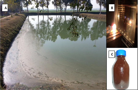
Sampling was performed from ponds and lagoons near wastewater treatment plant in Kasur. Sediment soil was collected from a region that was shallow enough to allow the reach of the sunlight and deep enough to be anoxic. Physical parameters such as pH and temperature were recorded. PNSB were isolated through enrichment culture technique by inoculating Mineral Salt Succinate (MSS) media bottles with the samples and adding a layer of mineral oil on the top for the development of anoxic conditions, followed by incubation at 30°C for one week under incandescent light (Figure 1) [15]. Isolated colonies were obtained by plating dilutions of concentration 10-2, 10-4 and 10-6 on MSS agar plates. The plates were incubated at 30°C under incandescent light for one week in an anerobic jar containing AnaeroGen sachet (Oxoid). The bacterial isolates were then characterized using microscopy and biochemical tests.
Figure 1. A: The sampling site. Samples were taken from ponds and lagoons near a wastewater treatment plant. B: Enrichment culture for PNSB under incandescent light. C: Enrichment of PNSB after the incubation
2.2 Toxic Metal Resistance
The bacterial isolates were tested for their ability to resist toxic heavy metals including As (Na3AsO4 and NaAsO2), Cu (Cu2SO4), Co (CoCl2), Cr (K₂CrO₄), Hg (HgCl2), Zn (ZnSO4), Ni (NiCl2), and Pb (PbCl2). MSS media petri plates, supplemented with heavy metals, were prepared. The bacterial isolates were cultured on these plates followed by an anoxic incubation of one week in anaerobic jar in the presence of incandescent light at 30°C.
2.3 Carbon Utilization Test
All the isolates were analyzed for the utilization of different carbon sources including succinate, citrate, acetate, oxalate, propionate and benzoate, with the final concentration of 10 Mm. MSS media tubes supplemented with each of these carbon sources were inoculated with 20 µl of overnight aerobic cultures of the bacterial isolates and incubated under both aerobic and anoxic conditions at 30°C. Continuous illumination by incandescent light was ensured in the case of anoxic incubation [16]. The bacterial isolates that showed growth aerobically but were unable to grow anaerobically were cultured again in the same media anaerobically with the addition of 5mM of As(V) to determine their ability to use the heavy metal as the terminal electron acceptor. The tubes were then incubated anaerobically at 30°C for one week under incandescent light [17].
2.4 Qualitative Assay of As Oxidation / Reduction
The ability of the bacterial isolates to reduce As(V) or oxidize As(III) was determined following the method reported by Simeonova, Lievremont, Lagarde, Muller, Groudeva and Lett [18]. MSS media tubes supplemented with either 1.0 mM of As(V) or As(III) were inoculated with overnight bacterial cultures and incubated at 30°C, on shaking at 100 rpm, for 48 hours. After the incubation, the cultures were centrifuged at 5000 rpm for 5 minutes, and supernatant was separated in fresh clean tubes. From each supernatant, 100 µl was transferred to micro-titer plate and mixed with 100 µl of 0.1 M silver nitrate solution. Positive and negative controls were also included alongside the samples. Development of yellow to brown colour was recorded as an indication of As reduction (change in colour from brown to yellow) or As oxidation (change in colour from yellow to brown).
2.5 Quantitative Assay of As Oxidation / Reduction
The extent of As oxidation / reduction by the PNSB was determined following the method reported by Cummings, Caccavo, Fendorf and Rosenzweig [19]. Overnight aerobic cultures incubated at 30°C were maintained at OD600 of 0.5 and were centrifuged for 5 minutes. The pellets were washed thrice with normal saline, and resuspended in 1.0 ml of phosphate buffer saline, 500 µl of which was added to 500 µl of 10 mM As(V) or 5 mM As(III) and incubated at room temperature for 48 hours. Samples were collected before and after the incubation, centrifuged, and the supernatant were transferred to fresh tubes. Supernatants were diluted with normal saline to achieve a concentration of 0.3mM for both As(V) and As(III).
100 µl of each supernatant was mixed with 100 µl of 24 mM HCl. 100 µl of the acidified samples were mixed with 900 µl of freshly prepared coloring reagent and vortexed immediately. The tubes were incubated at 78°C for 20 minutes, followed by incubation on ice for 5 minutes. Optical density was taken at 865nm using a spectrophotometer [19].
2.6 Determination of As(V) Reduction by the Bacterial Isolates in Artificial Wastewater
The ability of the bacteria to detoxify As was analyzed in artificial wastewater that consisted of (L-1): 1.0 g of sucrose, 0.2 g of NH4Cl, 0.15 g of CaCl2.2H2O, 0.33 g of KCl, 0.30 g of NaCl, 3.15 g of Mg2Cl, 1.26 g of K2HPO4, 0.42 g of KH2PO4, 1.0 g of yeast extract, and 1.0 ml of trace metals solution [20, 21]. Sterile artificial wastewater flasks supplemented with 5.0 mM of As(V) were inoculated with overnight cultures of the bacterial isolates. The flasks were incubated at 37°C for 48 hours at 120 rpm shaking. As(V) concentration was estimated following the incubation as given above.
2.7 Genomic DNA Isolation
Genomic DNA of the selected bacteria was isolated by CTAB method [22]. Bacteria were cultured aerobically at 30°C, and the cells were harvested by centrifugation. Pellets were suspended in 576µl Tris EDTA followed by addition of 30µl of 10%SDS and 3µl of 3M sodium acetate. The tubes were incubated for one hour at 37°C. 100µl of 5M NaCl was added to the tubes and mixed thoroughly followed by the addition of 80µl of CTAB-NaCl solution. The suspension was mixed thoroughly and incubated for 10 minutes at 65°C. One volume of chloroform:isoamyl alcohol (24:1) was added and centrifuged for 5 minutes at 12,000 rpm. Supernatants were transferred to fresh tubes followed by the addition of 0.6 volume isopropanol with gentle mixing. The tubes were centrifuged for 5 minutes at 12,000 rpm, and the pellets were washed with 70% ethanol. The pellets were air-dried and resuspended in 100µl TE buffer and stored at -20°C. DNA yield was confirmed through gel electrophoresis using 1.0% agarose gel.
2.8 Polymerase Chain Reaction and Phylogenetic Analysis
16S rRNA gene was amplified by using primers 27F (5-AGAGTTTGATCCTGGCTCAG-3) and 1522R (5-AAGGAGGTGTGCCARCCGCA-3) [23]. Amplification was carried out using thermal cycler (Advance Primus 96) with the following parameters: initial denaturation for 3 min at 94°C, followed by 30 cycles each consisting of denaturation for 45 sec at 94°C, annealing for 60 sec at 50°C, and extension for 90 sec at 72°C, followed by a final extension for 10 min at 72°C. PCR products were sequenced from Macrogen (Seoul, South Korea). The sequences were classified using NCBI BLAST tool (https://blast.ncbi.nlm. nih.gov/Blast.cgi). Top homologous sequences were downloaded and Neighbour Joining phylogenetic tree was made MEGA 5.0 [24] with a bootstrap value of 100 replicates [25].
2.9 Statistical Analysis
All the experiments were performed in triplicates or mentioned otherwise. The bars of the graphs represent mean values whereas the error bars represent standard deviations. The statistical analysis was performed in Microsoft Excel (Office 365).
3. RESULTS
3.1 Enrichment and Identification of As-resistant PNSB:
Purplish-brown colored bacterial colonies were obtained on MSS agar plates upon anoxic incubation in the presence of incandescent light (Figure 1). All the isolates were gram negative rods and showed positive results for catalase and oxidase tests. The bacterial isolate F1 was selected for identification using ribotyping based on the best overall results. 16SrRNA sequence analysis revealed the identity of the bacterial isolate as Rhodopseudomonas palustris, showing 99.63% identity with this bacterium in NCBI BLAST results. The identification was also confirmed through neighbour joining phylogenetic tree in which the bacterial isolate F1 grouped close to Rhodopseudomonas palustris (Figure 2).
3.2 Toxic Metal Resistance
All the isolates were resistant to Ni (1 mM), As(V) (2.5 mM), As(III) (1 mM) and Zn (1 mM) under both aerobic and anoxic conditions. Only R. palustris F1 showed resistance against Cr (0.1 mM) aerobically. All the bacteria were unable to resist As(III) (2.5 mM) anoxically. R. palustris F1 showed resistance against most of the heavy metals. None of the isolates showed resistance against Hg and Cu (Figure 3).
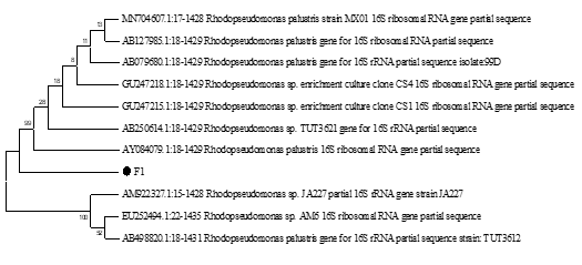
Figure 2. Neighbour joining phylogenetic tree of the bacterial isolate F1, constructed using MEGA 5.0 software. Bootstrap test values were used as measure of confidence, with values displayed at each node. Bacterial isolate F1 was found closely related to Rhodopseudomonas palustris
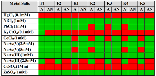
Figure 3. Resistance of PSNB against different toxic metals under both aerobic and anaerobic conditions
Table 1. Utilization of different Carbon sources by PNSB under aerobic and anaerobic conditions
|
Carbon Source Utilization Under Aerobic Conditions |
|||||||
|
Sr. No. |
Isolates |
Sodium Succinate |
Sodium Citrate |
Ammonium Oxalate |
Sodium Propionate |
Sodium Benzoate |
Sodium Acetate |
|
1 |
F1 |
+++ |
+ |
- |
+ |
- |
+ |
|
2 |
F2 |
+++ |
+ |
- |
+ |
- |
+ |
|
3 |
K1 |
+++ |
+ |
- |
+ |
- |
- |
|
4 |
K2 |
+++ |
+ |
+ |
+ |
- |
- |
|
5 |
K3 |
+++ |
+ |
- |
++ |
- |
+ |
|
6 |
K4 |
+++ |
+ |
- |
+ |
- |
- |
|
7 |
K5 |
+++ |
+ |
- |
++ |
- |
+ |
|
Carbon Source Utilization Under Anoxic Conditions |
|||||||
|
Sr. No. |
Isolates |
Sodium Succinate |
Sodium Citrate |
Ammonium Oxalate |
Sodium Propionate |
Sodium Benzoate |
Sodium Acetate |
|
1 |
F1 |
+++ |
- |
- |
- |
- |
- |
|
2 |
F2 |
+++ |
- |
- |
- |
- |
- |
|
3 |
K1 |
+++ |
+++ |
+ |
++ |
- |
- |
|
4 |
K2 |
+++ |
+++ |
+ |
++ |
- |
- |
|
5 |
K3 |
+++ |
- |
- |
- |
- |
- |
|
6 |
K4 |
+++ |
+++ |
+ |
++ |
- |
- |
|
7 |
K5 |
+++ |
- |
- |
- |
- |
- |
Table 2. Arsenic [As(V)] respiration by PNSB using different carbon sources under anoxic conditions
|
Sr. No. |
Isolates |
Sodium Succinate |
Sodium Citrate |
Sodium Propionate |
Sodium Benzoate |
Sodium Acetate |
|
1 |
F1 |
+++ |
+ |
+ |
|
- |
|
2 |
F2 |
+++ |
++ |
+ |
|
- |
|
3 |
K1 |
+++ |
|
|
- |
- |
|
4 |
K2 |
+++ |
|
|
- |
- |
|
5 |
K3 |
+++ |
++ |
+ |
|
- |
|
6 |
K4 |
+++ |
|
|
- |
- |
|
7 |
K5 |
+++ |
+ |
- |
|
- |
3.3 Carbon Utilization Test
Aerobically, visible growth of all the bacteria was observed in the case of sodium succinate, sodium propionate and sodium citrate. None of the isolates utilized sodium benzoate as a carbon source. Isolate K2 was the only isolate that showed growth in media containing ammonium oxalate, whereas isolates R. palustris F1, F2, K3 and K5 utilized sodium acetate as a carbon source. Anoxically, all the isolates showed growth for sodium succinate. K1, K2 and K4 showed growth for sodium citrate, ammonium oxalate and sodium propionate. None of the bacterial isolates were able to use sodium benzoate as a carbon source under anoxic conditions (Table 1).
The isolates were then cultured again anoxically in MSS media containing the carbon sources which they were unable to utilize under anoxic conditions and were supplemented with 5 mM As(V). R. palustris F1, F2, K3 and K5 showed growth in sodium citrate media supplemented with 5mM As(V), under anoxic conditions. R. palustris F1, F2 and K3 also showed growth in the presence of sodium propionate under anoxic conditions. This was an indication of their ability to use As(V) as a terminal electron acceptor under anoxic conditions (Table 2).
3.4 As Detoxification by the Bacterial Isolates
Initially, a colorimetric qualitative assay was performed to check the ability of the bacteria to oxidize / reduce As. R. palustris F1 was found to oxidize As(III), as development of brown colour was observed on the addition of the reagent, an indication of the presence of As(V). As(III) oxidation was also exhibited by K3 and K5. In the case of As(V) reduction, positive results were shown by all the isolates, as development of yellow colour was observed on the addition of the reagent, an indication of the presence of As(III). Most intense yellow colour development was observed in the case of the isolates K1, K4, K5 and R. palustris F1 (Figure 4).
The extent of As oxidation / reduction was also determined using a quantitative assay. Maximum As oxidation was observed in the case of R. palustris F1 which oxidized 4.31 mM of As(III). Isolates K4 and K5 oxidized As(III) up to 3.6 and 2.32 mM, respectively (Figure 5). In case of As reduction, the highest As(V) reduction was given by R. palustris F1, up to 7.46 mM. Bacterial isolate K5 and K4 reduced As(V) up to 7.15 and 6.38 mM, respectively (Figure 5). The ability of R. palustris F1 to reduce As(V) in artificial wastewater was also analyzed. It was found that R. palustris F1 reduced 3.6 mM of As(V) in artificial wastewater after 48 hours of incubation.
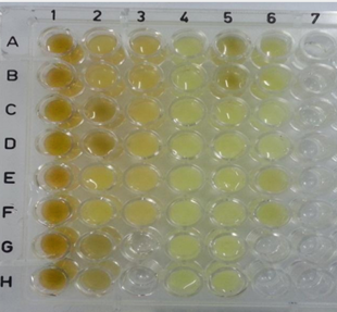
Figure 4. Qualitative test to determine the ability of the PNSB to oxidize / reduce As. Brown colour indicates the presence of As(V) whereas yellow colour indicates the presence of As(III) (Column 1: As(V) controls, Column 2: As(V) + bacterial samples F1, F2, K1 and K2 (in replicates), Column 3: As(V) + bacterial samples K3, K4 and K5 (in replicates), Column 4: As(III) controls, Column 5: As(III)+bacterial samples F1, F2, K1 and K2, Column 5: As(III)+bacterial samples K3, K4 and K5)
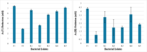
Figure 5. (A) As(V) reduction by PNSB. Maximum As(V) reduction was shown by Rhodopseudomonas palustris F1. (B) As(III) oxidation by PNSB. Maximum As(III) oxidation was shown by Rhodopseudomonas palustris F1
4. DISCUSSION
Microorganisms are known to play important roles in biotransformation and immobilization of toxic heavy metals in the environment, and thus find many biotechnological applications such as bioremediation [26]. Bacterial isolates in this study produced purplish-red-colored pigments when cultured anoxically in the presence of light, which is a characteristic of PNSB, and the colour is due to the pigments production such as chlorophyll and carotenoids. The purple / maroon colour shade varies depending on the exact composition and arrangement of these pigments in different PNSB [27]. The colour of the colonies, however, varied a bit, being pinkish red for K1 and F2, maroon or purplish red for K3 and K5 whereas brownish red for R. palustris F1. This variation could also may be attributed to the presence of different pigments lying in diverse absorption ranges [27,28]. Anaerobically, higher pigmentation is reported by bacteria grown in media supplemented with fructose [30]. Interestingly, the pigmentation ability is oxygen dependent. This reliance owes to the fact that the photosynthetic lamellar membrane, which harbors the carotenoids and chlorophyll, is induced under limiting oxygen conditions [31].
Microorganisms living in industrially polluted environments have adapted to the ability to resist and detoxify heavy metals. PNSB in this study were found to resist different heavy metals under both aerobic and anoxic conditions, showing their potential application to remediate industrially contaminated sites [32]. However, minor differences were found in the resistance observed under aerobic and anoxic conditions. None of the seven isolates showed growth against Hg and Cu, which could probably be due to the extremely toxic nature of these heavy metals. Moreover, the bacterial isolates may not have been directly exposed to these metals in the environment from where they were sampled, and thus might not have been adapted to resist such toxicants [33]. This relates to the complex, heterogenous nature of soil matrix, which contains many micro-sites. These micro-sites create localized environments, due to which compounds and nutrients are not uniformly available.
Although chemotrophic nutrition is a less favorable mode for bacterial growth; under conditions such as absence of incandescent light, facultative chemotrophic growth is an adaptation that enables the PNSB survive [34]. PNSB isolated in this study were able to utilize different carbon sources, both aerobically as well as anoxically, particularly sodium propionate, sodium citrate, and sodium succinate. These results supported the previously led studies [35]. R. palustris F1, F2, K3 and K5 utilized sodium acetate as their carbon source which indicates the presence of two key enzymes: isocitrate lyase and malate synthase. Both these enzymes are responsible for acetate assimilation via the citric acid cycle [36]. Anoxically, none of the bacterial isolates grew using sodium benzoate and sodium acetate as their carbon sources. The reason for inability to assimilate acetate could be the instability of the enzyme malate synthase, under anoxic conditions. Using oxygen as an electron acceptor, PNSB utilized these carbon sources as their source of energy, forming an electron donor-electron acceptor pair. This relationship, however, is disturbed under anoxic conditions. Therefore, for isolates that showed growth aerobically but failed to grow under anoxic conditions, the experiment was repeated with the addition of 5mM As(V). R. palustris F1, F2, K3 and K5 showed growth in sodium citrate media supplemented with 5mM As(V), under anoxic conditions. R. palustris F1, F2 and K3 also showed growth in the presence of sodium propionate under anoxic conditions. The results show that As(V) served as the terminal electron acceptor in the electron donor-acceptor reaction for these isolates under anoxic conditions. Thus, these bacteria were able to respire As anaerobically [37], a process known as dissimilatory As reduction. Such bacteria are also known as Dissimilatory arsenate reducing bacteria (DARB) [38].
PNSB in this study showed the ability of both oxidation as well as reduction of As in the qualitative assay, which was then confirmed quantitatively by a colorimetric assay. It was revealed that the bacterial isolates were able to reduce as well oxidize As. This is an indication of the presence of ars operon in these bacteria that confer the ability to resist and oxidize / reduce As [6, 39].
5. CONCLUSION
Such PNSB having the ability to bio-transform As via oxidation and reduction under both aerobic and anoxic conditions can find many biotechnological applications such as bioremediation, as these are ideal to work under a variety of conditions and even in the presence of other heavy metals which are normally found in industrially polluted sites.
Contribution of Authors
F.T.Z. performed the experiments, analyzed the data and prepared the original draft of the paper; Y.R. conceived the research, designed the experiments, analyzed the data and edited the final draft of the paper.
Conflicts of Interest
The authors declare no conflict of interest.
REFERENCES