Bioremediation of Textile Disperse Dyes using White-Rot Fungi Trametes gibbosa
Noor Fatima1 | Amina Nasir1 | Tehreem Hamid1 | Muhammad Akram1*
1Department of Life Sciences, School of Science, University of Management and Technology, Lahore, Pakistan
*Correspondence
Muhammad Akram
Email:
|
ABSTRACT The current study focuses on the biodegradation of disperse textile dyes and studies enzymes involved in degradation process. Various textile dyes are used in textile industry to dye different fabrics and polyesters. During the dyeing process, most of the dyes are not adsorbed and washed into the wastewater which affects the marine system and, ultimately, human health. Microorganisms like white rot fungi (WRF) have great potential of biodegradation of dyestuffs due to their active, nonspecific lignolytic catalytic systems. In this study, the white-rot fungi Trametes gibbosa was employed. The targeted disperse dyes were Disperse Blue-I and Disperse Red-I which are widely used in textile industries. The fungus were cultured on different media, and various biochemical parameters were optimized. Maximum growth of T. gibbosa was observed under optimized biochemical conditions (30 oC temperature, pH 6, 2% carbon source, 1% inoculum size and media peptone dextrose agar (PDA)). The decolonization process was optimized by using different concentrations of disperse dyes and wastewater. T. gibbosa show maximum degradation after 6 days incubation against disperse Red1 and disperse Blue1at the concentration of 0.02% (absorbance of 1.56 to 2.98), 0.01% (absorbance 0.22 to 2.87), respectively. The fungus showing maximum degradation at 2% wastewater concentration, with absorbance values ranging from 0.61 to 0.84. The addition of readily available carbon and nitrogen sources further enhanced the degradation efficiency. The overall efficiency of fungi was found to be more than 80%. The study concluded that T. gibbosa is a potential microorganism for the treatment of industrial effluents. The fungus also has potential for the production of active degrading enzymes such as laccase and lignin peroxidase. Further research is recommended to overexpress these enzymes, used purified enzymes to biodegrade diverse textile harmful and carcinogenic industrial effluents. Keywords: Bioremediation, Disperse dyes, Textile effluents, Trametes gibbosa, Wastewater |
Fungi have the remarkable ability to biodegrade pollutants and support a healthy environment. Due to its distinctive physiological traits, white-rot fungi (WRF) have the ability to rapidly degrade components of ecosystems [1]. White-rot fungi belong to basidiomycetes and, some ascomycete genera in the Xylariaceae family also exhibit white-rot properties. They produce extracellular enzymes responsible for breaking down lignin, cellulose, and hemicellulose [2]. Different environmental pollutants have been degraded inlcuding TNT (2, 4, 6-trinitrotoluene) by T. versicolor [3], pesticides [4] polycyclic aromatic hydrocarbons (PAHs) by Pleurotus spp.[5], P. chrysosporium [6], and T. versicolor. The Trametes genus comprises of white rot fungi found in various environments. In Europe, there are nearly nine species of this genus, which includes T. gibbosa, T. junipericola, T. cervina, T. ljubarskyi, T. ochracea, T. pubescens, T. hirsuta, T. suaveolens, and T. versicolor. T. gibbosa has pale-white fruiting bodies and light grey needle-like pores. These pores are ellipsoidal to sausage-shaped, and have a smooth surface [7]. It produces various enzymes such as laccase, manganese-dependent peroxidase, lignin peroxidase, glutathione S transferase and Cytochrome P450 (CYP450) [8][9].
Laccases are blue Cu²⁺ oxidases belonging to the family of oxidoreductases. Laccases are efficient at carrying out the oxidation of organic compounds such as polyphenols, monophenols, aromatic amines, and non-phenolic compounds by using oxygen [10]. Laccases oxidize a wide range of compounds while converting molecular oxygen into water [11]. Various isoforms of laccases in T. gibbosa have been identified depending upon the strains, locality, and environmental conditions. These include Laccase 1, Laccase 2, and Laccase A, B, D, F, and G. Laccase 1 and 2 comprise of only two plastocyanin-like domains, while Laccase A, B, D, F, and G consist of three such domains [12]. Lignin peroxidase (LiP) is encoded by MnP4 gene similar to Manganese peroxidase (MnP) in structure and function. MnP functions only with Mn²⁺ ions and their substrates specifically, while LiP can act on a diverse range of substrates [13]. Manganese peroxidase is a heme-containing peroxidase with a globular structure comprising approximately 11-12 α-helices. MnP degrades derivatives of lignin and xenobiotic compounds [13][14]. Four genes, i.e., Mnp1, Mnp2, Mnp3 and Mnp4, present in T. gibbosa, encode 364, 365, 366 and 362 amino acids, respectively, and share the same mechanisms of degradation [15]. Glutathione transferase (GST) transfers a glutathione molecule to electrophilic molecules and works with cytochrome P450 to degrade xenobiotics [8][16]. The substrates of GST are peroxides, for example, hydroperoxides of fatty acids, phospholipids, nitrate esters, etc. Cytochrome P450 proteins (CYPs) are a widespread protein family found in all domains of life. CYP450 is responsible in the discoloration of Congo red dye by P. chrysosporium [17].
Disperse dyes are synthetic textile dyes commonly used in colouring synthetic fibres, especially polyester. They are derived from chemical compounds, including azo, anthraquinone, and coumarin derivatives. Their low solubility in water makes them suitable for dyeing hydrophobic fibers like polyester [16][18]. Due to their hydrophobic nature, disperse dyes present environmental challenges by harming aquatic life and ecosystems. Thus, proper wastewater treatment and pollution control measures are essential to mitigate the environmental impact of disperse dyes and ensure their responsible use in the textile industry [18]. The use of dyes and pigments is increasing by 5.3% annually, in which textile industries are responsible for more than half of total dye consumption [19]. During the dyeing process in the textile industry, various salts and dyes are used, and around 10-15% are released into water bodies [20]. Approximately 2-20% of dyes are released into aquatic environments, and due to their persistence nature makes them unsuitable for aquatic animals’ consumption [21]. The persistent nature of dye adversely affects aquatic habitats, animals, and humans by causing ecotoxic, mutagenic, and carcinogenic effects [22]. To overcome these challenges, various chemical and physicochemical methods have been utilized. Different physical and chemical processes have been used for the treatment of industrial wastewater; however, they are not pocket and environment friendly [23]. Different biological methods, which are relatively simple and economical, have been the focus of current study for the dye biodegradation and decolorization [22][24]. The current study aims to develop a fast and inexpensive technique to treat textile wastewater effluent by using white rot fungus T. gibbosa. T. gibbosa has efficiency for the degradation of non-bioavailable substances like lignin, cellulose, and hemicellulose. Both standard dyes and wastewater from industrial effluent were used to evaluate the biodegradation ability of T. gibbosa.
This study explored the degradation of Disperse Red-1 and Disperse Blue-1 dyes using the fungus T. gibbosa. The T. gibbosa strain employed in the research was obtained from Azad Jammu Kashmir, Pakistan. It grows without a stalk on hardwood trunks and stalks of trees. Disperse Red 1 (DR1), Disperse Blue 1 (DB1) dyes were used as standards in this study. They were obtained in powdered form from Comfort Fabrics, Lahore, Pakistan. Wastewater from the textile industry was collected from Comfort Fabrics in Lahore, Pakistan, to be used as a growth medium for the fungi. Pure dye samples were utilized as references to determine and compare the concentration of dyes present in the wastewater samples.
The fungus (T. gibbosa) was identified by a lumpy surface, tough white flesh, and small needle-like pores on ventral surface, along with small spores about 4-5μm in size. T. gibbosa was purified and grown on both potato-dextrose agar and nutrient agar. Cultivation was carried out under mesophilic conditions, with a temperature range of 27–30 °C and a pH of 5–6, with a maximum incubation period of 3 days. To eliminate contamination, sub-culturing was performed multiple times. The purified culture was then transferred to a 50 mL broth medium and incubated at optimized conditions (Temperature 30 oC, pH 6) for 3 days to enhance the secretion of extracellular enzymes [25].
A 20% stock solution of each dye was prepared using Dimethyl Sulfoxide (DMSO) solution. Each flask contained 50 mL of broth in it. In total, 12 flasks were prepared: 1 blank flask having only broth with no dye or fungal culture, and 1 flask as a control with only fungal culture were added. The remaining 10 flasks were specified by concentrations of the dyes, i.e., 0.01%, 0.02%, 0.03%, 0.04%, and 0.05%. DR1 was added in 5 flasks, while DB1 in the other 5. The dilution formula was used to calculate the required volume of 20% stock solution as shown in Table 1:
C (stock) x V (stock) = C (dilution) x V (dilution)
Table 1. Volume of dyes used to prepare different stock dilutions
|
Dilution of Textile dye (%) |
Volume of Stock used (mL) |
|
0.01 |
0.0375 |
|
0.02 |
0.075 |
|
0.03 |
0.1125 |
|
0.04 |
0.150 |
|
0.05 |
0.1875 |
2.4 Biodegradation Assays with Dyes
From the fermentation culture, 3 mL samples were taken daily from day 1 to day 6 and analyzed both qualitatively and quantitatively. Qualitative analysis was done by visual examination of the color intensity for 6 continuous days, and the quantitative analysis was performed by using UV-Vis spectrophotometry. The absorbing wavelength used for analysis were 479 nm for DR1 and 615 nm for DB1. Negative control contained broth plus dyes (highest concentration being used in the experiments i.e. 0.05%.
Wastewater was diluted for effective the fungal degradation. The wastewater’s tentative composition contained different disperse dyes including orange, blue, red and black, but the specific concentrations of them were unknown. For the fungal cultures, 2% and 4% dilutions were used, which were calculated by:
C (stock) x V (stock) = C (dilution) x V (dilution)
Wastewater sample was added in the broth after 72 hours of growing the fungal cultures. Cultures were monitored by visual inspection and spectrophotometer assays were performed at daily intervals for up to 6 days. Positive and negative controls were also prepared following the same protocols used for dyes.
2.4.2 Biodegradation Assays in Wastewater
Wastewater collected from the textile industry contained Disperse Red, Orange, and Black dyes. The maximum absorbance values of Disperse Red and Orange dyes range between 470-500 nm (DR1 = 479 nm), However, black dye absorbance fall within 600-620 nm (DB-1 = 615 nm). Since the actual disperse dye composition was unknown, the average values of all possible kinds of dyes were calculated and applied to estimate spectrophotometric absorbance values. The absorbance values for disperse red and disperse orange dyes were observed at 480 nm, whereas 600 nm wavelength was used to measure Disperse Black dye. Broth in which the fungal cultures were grown was used as a blank. The graphs were plotted with every assay performed.
2.5 Optimization of Carbon and Nitrogen Sources
Statistical analysis was performed using 2way ANOVA and 1 sample t-test, by using SPSS software (version 19) for comparison between treatments. Significant differences between means of parameters were determined by using Duncan’s multiple range tests with probability ≤ 0.05.
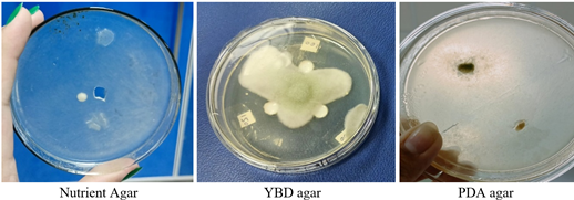
The fungal growth was optimized by growing it on different growth media. YBD, PDA, nutrient broth, and agar were used to culture this fungus. The biomass was observed on all plates by maintaining the temperature at30 oC and pH 6.
On PDA agar, the growth of both spores and mycelia was remarkably rapid, covering the entire plate within 72 hours. Due to rapid growth during sub-culturing, it became difficult to control the size of the inoculum, as even a small area contained a dense concentration of spores and mycelia. On YBD agar, despite the presence of two nitrogen sources, fungal growth was concentrated in a single spot on the plate. It produced a high number of spores while the mycelial biomass remained sparse. This also posed a challenge during subculturing, as the inoculum taken from the agar plates contained a high concentration of spores, resulting in rapid and exponential growth in the broth medium. On nutrient agar, the mycelial and spore biomass of T. gibbosa was not dense but rather spread across the plate. This medium was considered the most suitable nitrogen source, as it effectively regulated inoculum size, as shown in Figure 1.
Figure 1. Growth of T. gibbosa on different solid media
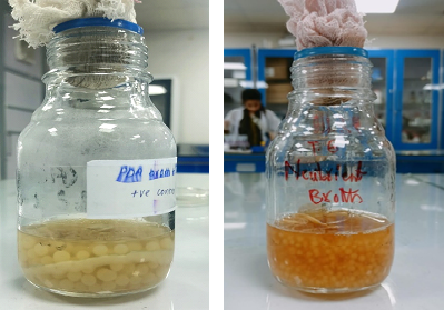
Figure 2. Growth of T. gibbosa on (a) PDA Broth (b) Nutrient Broth
Figure 3 shows the increase in biomass upon addition of nitrogen sources, keeping the carbohydrate source constant, i.e., glucose. The temperature and pH were also kept constant for this experiment. YBD medium presented the maximum growth as compared to all other media employed during experimentation.
The carbon sources for T. gibbosa were not optimized due to the unavailability of the necessary chemicals in the laboratory. Glucose was used as the carbon source in all experiments and showed acceptable performance across all media; nevertheless, the results remain inconclusive.
However, glucose was used as the carbon source in all experiments and showed acceptable performance across all media; nevertheless, the results remain inconclusive. Triplicate fungus culture was incubated at 25 oC, 28 oC, 30 oC and 37 oC while keeping the pH constant at 6. The growth duration was monitored to determine how quickly the fungal biomass increased and initiated spore formation. The appearance of spores indicates that the available nutrients in the medium have been consumed and the fungus has reached its maximum growth phase. Nutrient consumption varied with temperature. At 28 °C, the fungi were able to colonize the entire vessel within 72-96 hours. At 30 °C, the fungi were able to fully colonize the vessel in less than 72 hours. However, above the 30 °C temperature, fungal growth was significantly slower, taking up to 5 days to achieve a noticeable increase in biomass. At 37 °C, no fungal growth was observed, indicating that both mycelia and spores were unable to proliferate under normal bodily conditions. This observation supports the GRAS (Generally Recognized as Safe) status of T. gibbosa. The optimal pH for T. gibbosa is between 6.0 and 6.5. The experiments were conducted to check the growth at different pH values. A total of two batches of T. gibbosa were cultivated, one at the pH 6.0 and the other at pH 6.5.
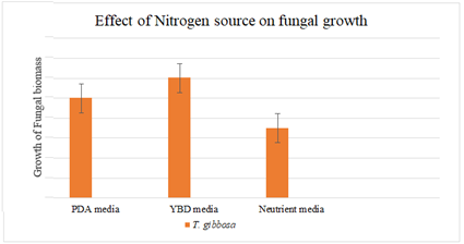
Figure 3. Effect of Nitrogen Source on Fungal Growth
The resulting curve (Figure 4) illustrates the rate of biomass growth over time, with the number of days represented on the x-axis. It depicts the progression of fungal development across the observed period. Starting from day 5, a decline in mycelial biomass was observed as spore formation began, which explains the drop in the growth rate between 96 and 108 hours shown in the curve (Figure 4).
After successful optimization of growth conditions, biodegradation experiments were carried out. The degradation rate was measured by UV-vis spectroscopy after every 24 hours. For this experiment, the fungal culture was incubated for 3 days at optimal conditions until the biomass had been produced. This step enabled the fungus to secrete extracellular ligninolytic enzymes into the medium, which would then be utilized for the degradation of the dyes. After 3 days of incubation, the dyes were added into the containers at varying concentrations prepared from a 20% stock solution. Five different dye concentrations were prepared, each with increasing levels of Disperse Red-1 and Disperse Blue-1. Spectrophotometric readings were recorded for both dyes at each concentration to assess their absorbance profiles. For red dye, maximum absorbance was recorded at 479 nm while for blue dye the value was 615 nm. These dyes were treated as standards in the experiments with wastewater.
3.5.1 Biodegradation by T. gibbosa
After the dyes were added into the medium, the spectrophotometric values were measured daily for 6 days. The results revealed a decline between 24 to 48 hours, followed by an increase with the passage of time. Although the dyes were visibly decolorized, the increase in absorbance values suggested that different intermediates were formed. This implied the production of other compounds, primarily intermediates of the degraded dye molecules. PDA broth used as a reference medium was taken for auto zero in the spectrophotometer at 479 nm.
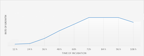
Figure 4. Growth Curve of T. gibbosa
|
P<0.05 |
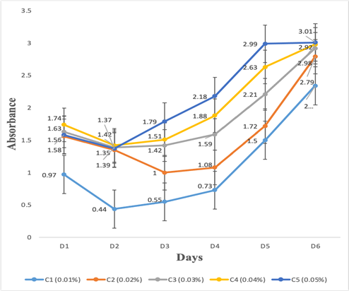
Figure 5. Degradation of Disperse Red-I by T. gibbosa
The value obtained was 0.30. This setup was also treated as a positive control, as the ideal outcome for dye degradation would result in complete breakdown of the dye, leaving the solution entirely clear. The value for negative control was calculated as 1.63 with the dye concentration of 0.05% solution, therefore it was similar to value of C5 on day 1. Any values between positive and negative controls were considered from day days 1 to 3. After that point, the absorbance values began to increase, due to intermediate production, making it difficult to compare the results with those of the positive and negative controls.
Figure 5 clearly shows that the degradation rate for T. gibbosa was extremely fast and degraded dye within 3 days of incubation. T. gibbosa successfully adsorbed all the dye molecules onto its mycelia within 12 hours of incubation. Following adsorption, the degradation of the dye molecules progressed gradually over the subsequent days. The results demonstrate there were significant effects on dyes bioremediation with respect to increase culture duration (days). Additionally, T. gibbosa was able to bind the intermediate compounds formed during the dye breakdown process. However, Figure 6 indicates some degree of ‘leakage’ of dye or its intermediates back into the medium, suggesting that not all compounds remained bound to the fungal biomass throughout the degradation process.
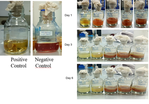
Figure 6. Visual Results of Degradation of Disperse Red-I by T. gibbosa over 6 days
|
P<0.05 |
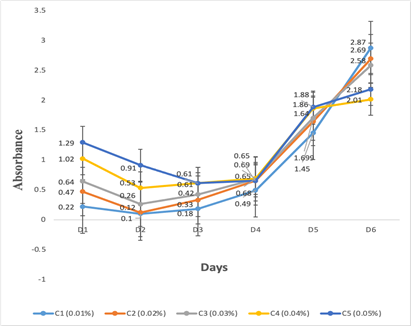
Figure 7. Degradation of Disperse Blue-I by T. gibbosa
Similar to DR1, a degradation curve was observed for D B-1. Tabular and graphical representation of degradation of DB-1 are shown in Figure 7. The absorbance values of broth were: negative control=1.32 and positive control=-0.20. A blank sample was used to auto zero the spectrophotometer. The results demonstrate there were significant effects on dyes bioremediation with respect to increase culture duration (days). The occurrence of negative values can be attributed to the limited solubility of dye molecules in the broth medium, which prevented a measurable increase in absorbance. Additionally, as the degradation process progressed over time, the values continued to decrease, becoming increasingly negative—likely due to further breakdown or removal of dye molecules from the solution. This was because the nutrients continued depleting from the growth media.
A visual representation of the degradation of DB1 during 6 days is shown in Figure 8. The visual results confirmed that the dyes were being degraded, unlike the absorbance values. An increase in absorbance values may be due to the degrading ring structure in the dye molecule, which gives them more exposure to absorb light photons coming from the spectrophotometer.
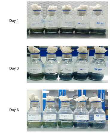
Figure 8. Visual Results of Degradation of Disperse Blue-I by T. gibbosa
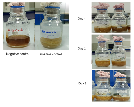
Figure 9. Biodegradation of textile wastewater by T. gibbosa over 3 days
Experiments with wastewater demonstrated that T. gibbosa was effective in degrading the wastewater within the 3 days of incubation, as shown below in Figure 9.
|
P<0.05 |
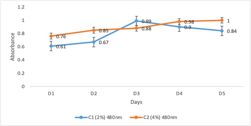
Figure 10. Degradation of Disperse Red and Orange by T. gibbosa
|
P<0.05 |
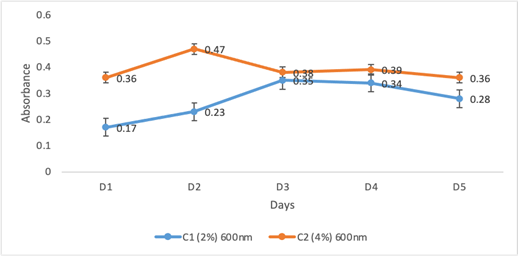
Figure 11. Degradation of Disperse Black by T. gibbosa
The degradation rate was monitored by using spectrophotometer. The absorbance values were in accordance with the degradation results of DR1 and DB1 in the broth. As the degradation process progressed over time, the values continue to decrease, becoming increasingly negative likely. This was because the medium nutrients kept on depleting from the growth medium. The figure clearly shows the absorbance values of wastewater at different wavelengths (Figure 10 and 11). Two graphs were drawn; one for the wavelength of disperse orange and disperse Red, which lies between 480 nm (Figure 10) and the other for disperse black dye which absorb around 600 nm (Figure 11). The degradation rate was faster in the 2% dye solution, as shown in Figure 10 and 11. The results demonstrate there were significant effects on wastewater dyes bioremediation with respect to increase culture duration (days).
Yaseen and Scholaz reported the characteristics of textile industry wastewater effluent and its impact on microbial and aquatic life [20]. WRF enzymes are able to degrade textile disperse dyes. The focus of this research was first to optimize the growth conditions of T. gibbosa and then assessing its ability to degrade disperse dye containing wastewater. Several studies in the past on T. gibbosa have assessed the activity of biodegradation on various substrates, but enzymatic activators and surfactants had been used to enhance the activity of enzymes [8][25][26]. After optimization of T. gibbosa, the best growth was observed at 30 oC. Adnan et al. and Knežević et al. reported the optimized growth of T. gibbosa at an optimum pH 5 and temperature of 25 oC. Hence, the growth conditions are different in this study than Adnan et al [26]. However, Knežević et al. conducted an experiment to degrade wheat straws using a wild type T. gibbosa, while the cultures were grown on Malt agar with temperature 22 ± 2 oC for 7 days [27]. In our study, the growth obtained was within 3 days of incubation at 30 oC. For this reason, 30 oC is more efficient than room temperature as used in Knežević’s research [27]. The optimum pH observed was 5.5-6.0 but a pH 6 is considered optimal. Therefore, the optimum pH varies in accordance with the specific purpose of the research [28][29]. Previously, this study used Malt agar as a growth medium for T. gibbosa. Malt extract is rich in various carbohydrates, including sugars such as glucose, maltose, and other complex carbohydrates. In our experiments, the pH was maintained at 6 for T. gibbosa, which has been evidenced to be the most optimal in previously published literature [30]. In the past, T. gibbosa has been used to biodegrade a few compounds, including its natural substrates and pollutants. Further research is required in the assessment of the T. gibbosa’s ability to biodegrade pollutants due to its potential in the field of environmental biotechnology. Eshghi et al. demonstrated that T. gibbosa efficiently decolorize (more than 80%) the textile dyes Methyl Green (MG), Methylene Blue (MB) and Reactive Black 5 (RB5). This efficiency was due the enzyme MnP by adding MnP activator and maximum time period for culture (30 days) [25]. In our study, when samples were incubated for more than 8-9 days, the fungal culture became so dense that produced different intermediates; which interfered with the degradation results. Therefore, the accurate results that have been measured were for 6 days. The nature and chemical structure of the intermediates could not be examined due to no availability of GC-MS or FTIR, so that we could have identified the types of bonds attached on by fungal enzymes. The current study has different limitation as using controlled laboratory conditions, only disperse textile dyes were targeted, used whole culture for bioremediation and fungus was exposed to dyes and wastewater for a short period of time. The limitations can be overcome by scaling up the process to pilot or industrial level; can use other types of dyes for decolonization and using the advance level molecular techniques to overexpress the lignolytic enzymes (laccase and lignin peroxidase) that might contribute to bioremediation. Exploring the synergistic effects of T. gibbosa in combination with other fungi or bacteria could lead to more robust and efficient bioremediation systems. Further optimization of culture conditions (pH, temperature, aeration, and carbon/nitrogen ratios), could significantly enhance fungal growth and enzymatic activity.
The research focused on the ability of WRF, particularly T. gibbosa, to bioremediate wastewater by degrading disperses textile dyes. T. gibbosa was cultured on different agar and broth media. The incubation period and a number of growth parameters, including pH, temperature, carbon and nitrogen supplies, were optimized to enhance the production of fungal biomass and enzymes. T. gibbosa had the ability to degrade Disperse Red-I and Blue-I dyes as well as other dyes commonly present in industrial effluent. In particular, T. gibbosa have a high dye degradation efficiency which was confirmed by the spectrophotometric analyses. The results show that wastewater contaminated with textile dyes can be treated effectively and sustainably by employing white-rot fungi. The study concludes that T. gibbosa has strong potential to degrade disperse dyes by over 80%, making it a promising candidate for treating industrial effluent. Further studies could also investigate its effectiveness in degrading other toxic types of dyes and industrial wastewater.
Funding
This research received no external funding.
Acknowledgements
This study was supported by the Department of Life Science, University of Management and Technology, Lahore, Pakistan.
Conflicts of Interest
The authors declare no conflict of interest.