Isolation and Characterization of Antibiotic-producing Bacteria from the Salt Range of Kallar Kahar, Pakistan
Noreen Iftikhar[1]*, Iqra Khalid[2]
KEYWORDS
Agar plug method, Antibiotics, Starch-casein agar, Streptomyces
The emergence of antibiotic resistance in pathogenic bacteria has heightened the need for new antibiotics. Streptomycetes are filamentous gram-positive bacteria that are ubiquitous and present in saline soil and produce antibiotics as secondary metabolites under stressful conditions. Naturally, Streptomyces produce over two-thirds of antibiotics that are used clinically. Saline soil was collected from Kallar Kahar, Pakistan. The soil sample was serially diluted and three dilutions were plated on Streptomyces selection media (starch-casein agar and glucose yeast malt agar) after growing the culture, pure colonies were selected based on their morphological features and subsequently examined using Gram-staining. The antimicrobial activity of two selected strains (P1 and P2) was evaluated using the agar plug method and agar well diffusion method against both gram-positive (Bacillus subtilis) and gram-negative (Escherichia coli) pathogenic bacteria. In the agar plug method, the clear zone of inhibitions was not clearly visible against the test bacteria. The zone of inhibitions were only observed in agar well diffusion assay in which the P1 strain exhibited a diameter of 0.6mm against E. coli and 0.75mm against B. subtilis and the P2 strain showed antibacterial activity only against E. coli with a diameter of 0.75mm. The results were not significant, these slight zones of inhibition warrant further improvements in methods for isolation and purification of antibiotic-producing bacteria. Such methods should aim to enhance the efficiency of antibiotics.
An antibiotic is derived from the word ‘antibiosis’ which means ‘against life’. In the initial days, antibiotics were considered to be organic compounds that are produced by microbes. This initially defines the concept of antibiotic as a substance produced by microbes that inhibit the growth or kill pathogenic microbes. However, the definition has been expanded in recent times to include the synthetic compounds that mimic the properties of natural antibiotics [1]. Initially the term antibiotic was used to describe the formulations of living organisms but now this term has been extended to encompass a broader range to many other antimicrobials that produced synthetically such as fluoroquinolone and sulphonamides [2]. Synthetic antibacterial drugs are used for the treatment but their use can reduce the amount beneficial gut flora, leading to adverse health effects. This reduction in gut flora decrease the efficiency of other drugs against bacterial infections. Several health problems have been observed in patients who used antibacterials for the long term [3].
Natural products are more readily available due to their production by living organisms. Antibiotics derived from natural sources are employed in the treatment of bacterial infections [4]. These natural products are produced by living organisms as a defense mechanism against predators, aiding the organism’s adaption to the environment. Through the process of natural selection, these products have evolved to endure various biotic and abiotic stresses [5]. Antibiotics can be classified as microbicidal or microbiostatic, the former kills the bacteria or the later inhibits the bacterial growth. Antibiotics exhibit a variety of modes of action, including inhibiting enzymes regulations, disrupting metabolic reactions, interfering with cell wall and cell membrane formation and functions, and blocking nucleic acids and proteins synthesis [6]. Antibiotics are classified based ontheir spectrum of mechanism of action either broad or narrow. Broad-spectrum antibiotics can inhibit or kill a broad range of microbes while narrow-spectrum antibiotics can only kill or inhibit a specific group of microbes.
With the growth of the human population, the emergence of bacterial infections has intensified claiming millions of lives, particularly in developing countries. In Bangladesh, Pakistan, and India only infectious diseases have caused 3.7 million deaths [7]. The emergence of antibiotic resistance in pathogenic bacteria has created urgent need for new antibiotics. The first antibiotic, penicillin, was discovered naturally from the fungus Penicillium notatum [8]. Many other antibiotics used for clinical purposes such as erythromycin, chloramphenicol, neomycin, tetracycline, streptomycin, and gentamycin are derived from actinomycetes, gram-positive, filamentous, and soil-dwelling bacteria [9]. These belong to genus Streptomyces, which produces antibiotics as secondary metabolites that are approved for human use. Due to the presence of filaments, Streptomycetes comprise approximately 40% of soil bacteria. These organisms require only inorganic nutrients for growth and contribute to an impressive 80% of all antibacterial drugs [10]. This study focuses on screening antibiotic-producing microbes from salt-rich areas, as actinomycetes are more abundant in saline soil. These actinomycetes have potential to produce antibiotics that could be used against bacterial infection. The increasing prevalence of antibiotic resistance so underscores the need to discover novel antibiotics that can effectively combat the pathogenic bacteria.
Chemicals and Reagents
All the chemicals and reagents were procured from Sigma-Aldrich, while agar and starch were purchased from Merck. Bacterial strains were sourced from the University of Management and Technology, Lahore, Pakistan. These strains were employed to evaluate the antibacterial activity of Streptomyces isolates. Two strains were utilized, one was Gram-positive (Bacillus subtilis) and the other was Gram-negative (Escherichia coli). Both strains were maintained on nutrient agar slants at 4˚C or in 20%v/v glycerol at -20˚C.
Sample Collection and Sites
Six soil samples were collected from various salt-rich areas in Kallar Kahar, Pakistan. The Using sterile spatulas, samples were collected and transferred to the aseptic bag, and transported to the laboratory of Department of Life Sciences, University of Management and Technology, Lahore, Pakistan for further processing.
Streptomyces Selection Media and Plating of Serial Dilutions
Starch-casein agar (SCA) medium was employed for the growth of bacterial colonies. The composition of SCA media is as follows: (10g starch, 0.3g casein, 2g KNO3, 2g K2HPO4, 2g NaCl, 0.02g CaCO3, 0.01g FeSO4.7H2O, 0.05g MgSO4.7H2O, 15g agar, 1000ml distilled water and having pH:7). Streptomyces colonies were selectively isolated using glucose yeast malt extract (GYM) agar media, which has the following composition: 5g yeast extract, 5g malt extract, 10g glucose, 15g agar, and 1000ml distilled water [11]. To prepare the serial dilutions, 1g of soil was added to 10ml of saline solution and the suspension was vortexed for 45 seconds. Different serial dilutions (1:10, 1:100, and 1:1000) were made in a total volume of saline solution of 10ml. 50µl of each serially diluted samples were spread on starch-casein agar and the sample plates were incubated at 28˚C for 7-21 days to allow bacterial growth.
Isolation of Pure Streptomyces Colonies
Colonies exhibiting morphological features characteristics of Streptomyces were selected and using sterile loops and inoculated onto fresh SCA plates to confirm their purity. The colonies were then transferred to GYM agar and incubated at 28˚C for 3 days to facilitate further morphological characterization. Observations were made by regarding the color of substrate mycelium, growth patterns, pigment production, and spore-mass color. Gram staining was performed on pure colonies, followed by examination under a light microscope. Sub-cultures were prepared by innoculating 250ml of GYM broth with the colonies and incubating at 28˚C for three days at 150rpm.
Antimicrobial Assay
Assays were conducted to evaluate the inhibitory effects of secondary metabolites against microbes and to determine the efficiency of antibiotics. Antimicrobial activity was assesssed using solid media bioassays against Gram-positive and Gram-negative bacteria. The two bioassays employed are described below: the agar plug method and agar well diffusion method.
Agar Plug Method
In the agar plug method, isolated strains were densely tightly onto the surface of GYM agar plates. During incubation at 28˚C for seven days, microbial cells secreted secondary metabolites that diffused into the medium. After incubation, a cylinder or agar plug was cut using a cork borer and placed onto the surface of another agar plate that had been inoculated with test microbes. The antibacterial activity of the diffused molecules was detected by measuring the zone of inhibitions around the plug.
Agar Well Diffusion Method
This procedure involves inoculating the entire surface of agar plates with bacterial strains by spreading the inoculum. Next, wells with diameter 6-8mm were created in the agar plates usin sterile blue tips or cork borer, and a solution of the microbial agent is added to each well. The agar plates were then incubated at 37˚C for 24 hours, after which the diameter of the zone of inhibition around each well was measured.
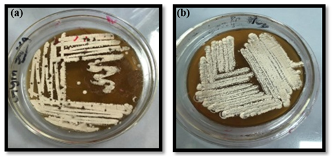
Figure 1. Two strains purified from the soil sample (a) P1 strain (b) P2 strain
Selection of Pure Colonies
The colonies were selected based on their morphological characteristics. The purified strains were creamy or yellowish in color with thread like filaments that appeared spiny and extended outwards to the peripheral surface. These filaments were long and straight, containing numerous spores (>50) arranged in whorls. The mycelium was present and had well-developed hyphae with multiple branches. The two strains of the genus Streptomyces were purified, as shown in Figure 1.
Gram Staining of Pure Cultures
The pure cultures were stained using Gram staining to determine whether they are gram-positive or gram-negative. Upon staining, the cultures were examined under a light microscope, confirming that the strains were gram-positive which and belonged to the genus Streptomyces. The stained culture is shown in Figure 2.
Agar Plug Method Against Test Bacteria
The molecules from the plug diffused into the agar plates, allowing the antibacterial activity of those molecules against test bacteria to be determined. The zone of inhibitions around the plug confirmed the antibacterial activity of diffused molecules. However, the zone of inhibitions around the plug was not clearly visible for both strains, as shown in Figure 3.
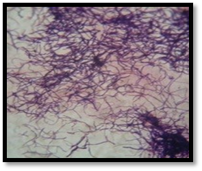
Figure 2. Gram staining of pure-culture confirmed them to be gram-positive
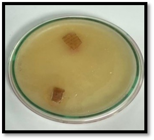
Figure 3. No clear zone of inhibitions was observed in the agar plug method
Agar Well Diffusion Against Test Bacteria
The antibacterial activity of the selected bacterial isolates was determined using the agar well diffusion method. The P1 strain demonstrated a larger zone of inhibition against both test bacterial strains than the P2 strain. The P1 strain inhibited the Gram-positive bacteria Bacillus subtilis with a 0.75mm distance of inhibition (DOI), while the P2 strain did not inhibit the Bacillus subtilis as evidenced by no zone of inhibition. The P1 also exhibited a clear zone of inhibition against Gram-negative bacteria Escherichia coli measuring a 0.6mm in diameter but the P2 strain exhibited a larger inhibition zone against Escherichia coli, measuring 0.75mm in diameter. A comparison of the inhibitions zones of both strains against both test bacteria is presented in Figure 5 and Figure 4.
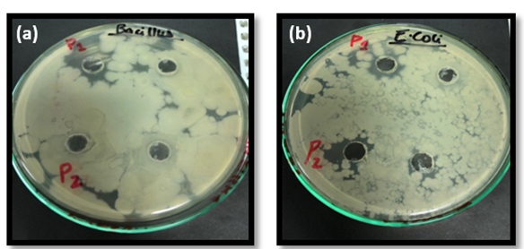
Figure 4. Agar well diffusion: Antibacterial activity of Streptomyces extract against (a) B. subtilis (b) E. coli

Figure 5. Diameter of zones of inhibition of P1 and P2 strain against Bacillus subtilis and Escherichia coli
Antibiotic resistance among pathogenic bacteria has been on rise, leading to the ineffectiveness of existing antibiotics in treating infections. This highlights the increased needto discover new antibiotics, particularly from natural sources [12]. Among various methods, the Waksman platform has emerged as most effective approach for screening the antibiotic-producing microbes from the soil [13]. The main purpose of this study is to isolate antibiotic-producing bacteria from saline soil, as soil inhabitants are predominantly represented by the genus Streptomyces. These filamentous bacteria are rich source of natural bioactive compounds with diverse biological activities, making them valuable for pharmaceuticals and agrochemicals applications. Notably, Streptomycetes have contributed to the production of substantial number of compounds, with 75% of antibiotics originating from this genus [14]. This study investigated the isolations, selection process, and antimicrobial activities of the molecules produced by the Streptomyces.
Streptomyces’ filamentous structure, which provides resilience against the environmental stresses, are predominantly in dry and saline environments. Their filaments provide them strength to the saline soil texture that protects them from different environmental stresses [15]. A positive correlation between saline soil depth and presence of Streptomyces sp. as salinity exerts a significant influence on the bacterial composition of soil communities [16]. Streptomyces are aerobic, chemoorganotrophic bacteria that do not requ growth factors or vitamins to grow. The alkaline environment of saline soil, facilitates the bacterial isolation from those that are unable to tolerate salinity, and leaving only salt tolerating filamentous bacteria producing secondary metabolites under stressful conditions [17]. The exposure of extreme conditions as heat shock, ethanol treatment, and high hydrostatic pressures is a common strategy to produce bioactive compounds [18].
The diameter of the zone of inhibition is correlated to the antimicrobial activity of the bioactive molecule. A larger the zone of inhibition indicates the more efficient antimicrobial activity of molecules [19]. In the agar plug method, the zone of inhibitions has not been observed due to absence of effective concentration of compounds in the agar to inhibit bacterial growth. In the agar well diffusion method, the extracts from Streptomyces strains P1 demonstrates more efficacy than P2 as P1 inhibited the growth of both tested bacteria, while P2 was only effective against Escherichia coli. Slight zones of inhibition were observed around the wells by both isolated bacterial strains. However, the antimicrobial efficacy of antibiotic-producing strains was relatively low as these have higher minimal inhibitory concentrations [20]. There is a need to refine the techniques for isolating bacterial strains for the effective production of antibiotics and develop methods to increase their antimicrobial efficiency.
Naturally produced antibiotics offer several advantages chemically synthesized drugs. Antibiotic-producing microbes belong to the genus Streptomyces and are often found in saline environments. Isolated Streptomyces strains (P1 and P2) obtained from saline soil exhibited antimicrobial activity against the tested bacteria. Screening for novel antibiotic-producing bacteria can help in discovery of new drugs capable to combat pathogenic bacteria. The development of noval antibiotics is crucial to address the growing issue of antibiotic resistance. Further studies should involve DNA sequencing to confirm the bacterial strains at specie level. Additionally, methods have to enhance the efficacy of these antibiotics should be explored.
*Correspondence: noreeniftikhar0505@gmail.com
[1]Center of Excellence in Molecular Biology, University of the Punjab, Lahore, Pakistan
[2]Department of Life Sciences, University of Management and Technology, Lahore, Pakistan